1AD8

COMPLEX OF THROMBIN WITH AND INHIBITOR CONTAINING A NOVEL P1 MOIETY


Experimental Data Snapshot

  • Method: X-RAY DIFFRACTION
  • Resolution: 2.00 Å
  • R-Value Work: 0.205 
  • R-Value Observed: 0.205 

wwPDB Validation   3D Report Full Report


Ligand Structure Quality Assessment 


This is version 1.3 of the entry. See complete history


Literature

Molecular design and characterization of an alpha-thrombin inhibitor containing a novel P1 moiety.

Malikayil, J.A.Burkhart, J.P.Schreuder, H.A.Broersma Jr., R.J.Tardif, C.Kutcher 3rd., L.W.Mehdi, S.Schatzman, G.L.Neises, B.Peet, N.P.

(1997) Biochemistry 36: 1034-1040

  • DOI: https://doi.org/10.1021/bi9622231
  • Primary Citation of Related Structures:  
    1AD8

  • PubMed Abstract: 

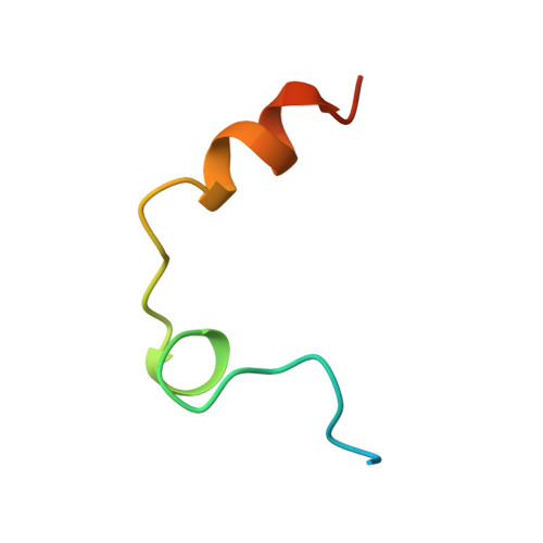
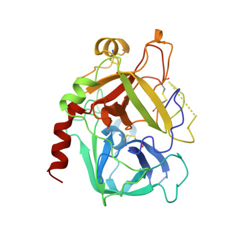
    An inhibitor of alpha-thrombin was designed on the basis of the X-ray crystal structures of thrombin and trypsin. The design strategy employed the geometric and electrostatic differences between the specificity pockets of the two enzymes. These differences arise due to the replacement of Ser 190 in trypsin by Ala 190 in thrombin. The new inhibitor contained a tryptophan side chain instead of the arginine side chain that is present in the prototypical thrombin inhibitors. This inhibitor had a Ki value of 0.25 microM, displayed more than 400-fold specificity for thrombin over trypsin, and doubled the rat plasma APTT at a concentration of 44.9 microM. The X-ray crystal structure of the inhibitor/alpha-thrombin complex was determined. This represents the first reported three-dimensional structure of a thrombin/ inhibitor complex where the specificity pocket of the enzyme is occupied by a chemical moiety other than a guanidino or an amidino group. As was predicted by the molecular model, the tryptophan side chain docks into the specificity pocket of the enzyme. This finding is in contrast with the indole binding region of thrombin reported earlier [Berliner, L. J., & Shen, Y. Y. L. (1977) Biochemistry 16, 4622-4626]. The lower binding affinity of the new inhibitor for trypsin, compared to that for thrombin, appears to be due to (i) the extra energy required to deform the smaller specificity pocket of trypsin to accommodate the bulky indole group and (ii) the favorable electrostatic interactions of the indole group with the more hydrophobic specificity pocket of thrombin. The neutral indole group may be of pharmacological significance because the severe hypotension and respiratory distress observed following the administration of some thrombin inhibitors have been linked to the positively charged guanidino or amidino functionalities.


  • Organizational Affiliation

    Hoechst Marion Roussel, Inc., Cincinnati, Ohio 45215, USA.


Macromolecules
Find similar proteins by:  (by identity cutoff)  |  3D Structure
Entity ID: 1
MoleculeChains Sequence LengthOrganismDetailsImage
THROMBIN (SMALL SUBUNIT)A [auth L]36Homo sapiensMutation(s): 0 
EC: 3.4.21.5
UniProt & NIH Common Fund Data Resources
Find proteins for P00734 (Homo sapiens)
Explore P00734 
Go to UniProtKB:  P00734
PHAROS:  P00734
GTEx:  ENSG00000180210 
Entity Groups  
Sequence Clusters30% Identity50% Identity70% Identity90% Identity95% Identity100% Identity
UniProt GroupP00734
Sequence Annotations
Expand
  • Reference Sequence
Find similar proteins by:  (by identity cutoff)  |  3D Structure
Entity ID: 2
MoleculeChains Sequence LengthOrganismDetailsImage
THROMBIN (LARGE SUBUNIT)B [auth H]259Homo sapiensMutation(s): 0 
EC: 3.4.21.5
UniProt & NIH Common Fund Data Resources
Find proteins for P00734 (Homo sapiens)
Explore P00734 
Go to UniProtKB:  P00734
PHAROS:  P00734
GTEx:  ENSG00000180210 
Entity Groups  
Sequence Clusters30% Identity50% Identity70% Identity90% Identity95% Identity100% Identity
UniProt GroupP00734
Sequence Annotations
Expand
  • Reference Sequence

Find similar proteins by:  Sequence   |   3D Structure  

Entity ID: 3
MoleculeChains Sequence LengthOrganismDetailsImage
HIRUDIN (53-65) PEPTIDEC [auth I]10Hirudo medicinalisMutation(s): 0 
UniProt
Find proteins for P01050 (Hirudo medicinalis)
Explore P01050 
Go to UniProtKB:  P01050
Entity Groups  
Sequence Clusters30% Identity50% Identity70% Identity90% Identity95% Identity100% Identity
UniProt GroupP01050
Sequence Annotations
Expand
  • Reference Sequence
Small Molecules
Ligands 2 Unique
IDChains Name / Formula / InChI Key2D Diagram3D Interactions
MDL
Query on MDL

Download Ideal Coordinates CCD File 
E [auth H][DEHYDROXY-N-METHYL-TYROSYL-PROLINYL]-[4,4,5,5,5-PENTAFLUORO-3-OXY-1-[3-INDOLYL]-PENT-2-YL]AMINE
C28 H29 F5 N4 O3
WFJABZVISMTOSO-ZRBLBEILSA-N
NA
Query on NA

Download Ideal Coordinates CCD File 
D [auth H]SODIUM ION
Na
FKNQFGJONOIPTF-UHFFFAOYSA-N
Modified Residues  1 Unique
IDChains TypeFormula2D DiagramParent
TYS
Query on TYS
C [auth I]L-PEPTIDE LINKINGC9 H11 N O6 STYR
Binding Affinity Annotations 
IDSourceBinding Affinity
MDL PDBBind:  1AD8 Ki: 250 (nM) from 1 assay(s)
Experimental Data & Validation

Experimental Data

  • Method: X-RAY DIFFRACTION
  • Resolution: 2.00 Å
  • R-Value Work: 0.205 
  • R-Value Observed: 0.205 
  • Space Group: C 1 2 1
Unit Cell:
Length ( Å )Angle ( ˚ )
a = 70.55α = 90
b = 72.05β = 101
c = 73γ = 90
Software Package:
Software NamePurpose
XDSdata scaling
XSCALEdata scaling
X-PLORmodel building
X-PLORrefinement
XDSdata reduction
X-PLORphasing

Structure Validation

View Full Validation Report


Ligand Structure Quality Assessment 


Entry History 

Deposition Data

Revision History  (Full details and data files)

  • Version 1.0: 1997-11-12
    Type: Initial release
  • Version 1.1: 2008-03-03
    Changes: Version format compliance
  • Version 1.2: 2011-07-13
    Changes: Atomic model, Database references, Derived calculations, Non-polymer description, Structure summary, Version format compliance
  • Version 1.3: 2024-04-03
    Changes: Data collection, Database references, Derived calculations, Refinement description