1ABJ

STRUCTURE OF THE HIRULOG 3-THROMBIN COMPLEX AND NATURE OF THE S' SUBSITES OF SUBSTRATES AND INHIBITORS


Experimental Data Snapshot

  • Method: X-RAY DIFFRACTION
  • Resolution: 2.40 Å
  • R-Value Observed: 0.144 

wwPDB Validation   3D Report Full Report


This is version 1.3 of the entry. See complete history


Literature

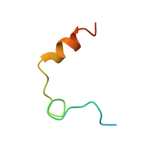
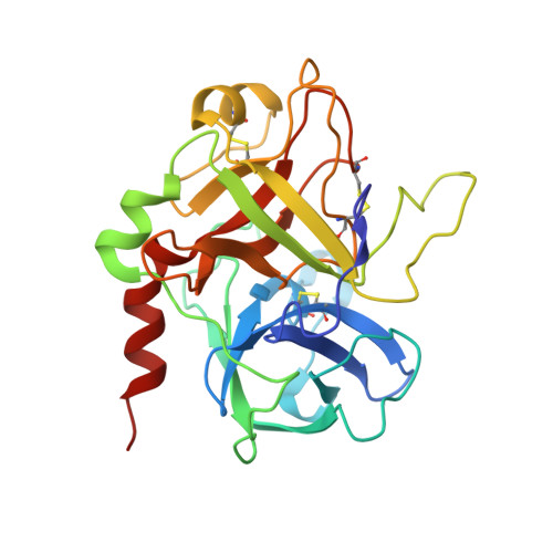
Structure of the hirulog 3-thrombin complex and nature of the S' subsites of substrates and inhibitors.

Qiu, X.Padmanabhan, K.P.Carperos, V.E.Tulinsky, A.Kline, T.Maraganore, J.M.Fenton 2nd., J.W.

(1992) Biochemistry 31: 11689-11697

  • DOI: https://doi.org/10.1021/bi00162a004
  • Primary Citation of Related Structures:  
    1ABI, 1ABJ

  • PubMed Abstract: 

    The X-ray crystallographic structure of the human alpha-thrombin complex with hirulog 3 (a potent, noncleavable hirudin-based peptide of the "hirulog" class containing a beta-homoarginine at the scissile bond), which is isomorphous with that of the hirugen-thrombin crystal structure, was solved at 2.3-A resolution by starting with a model for thrombin derived from the hirugen-thrombin complex and was refined by restrained least squares methods (R = 0.132). Residues of hirulog 3 were well-defined in the electron density, which included most of the pentaglycine linker and the C-terminal helical turn that was disordered in a related structure of thrombin with hirulog 1. The interactions of D-Phe1'-Pro2'-beta-homoArg3' with the active site of thrombin were essentially identical to those of related structures of PPACK- (D-Phe-Pro-Arg chloromethyl ketone) and hirulog 1-thrombin, with the guanidinium function of the arginyl P1 residue forming a hydrogen-bonding ion pair with Asp189 of the S1 site. A noticeable shift in the CA atom of beta-homoArg3' due to the methylene insertion displaces the scissile bond from attack by Ser195, thus imparting proteolytic stability to the beta-homoArg hirulog derivative. Resolution of the pentaglycine spacer, linking N- and C-terminal functional domains into a single oligopeptide bivalent inhibitor, permitted delineation of corresponding S' subsites of thrombin. The position of Gly4' (P1') is stabilized by three hydrogen bonds with His57, Lys60F, and Ser195, while the conformational angles maintained in a strained, nonallowed configuration for non-glycyl amino acids.(ABSTRACT TRUNCATED AT 250 WORDS)


  • Organizational Affiliation

    Department of Chemistry, Michigan State University, East Lansing 48824-1322.


Macromolecules
Find similar proteins by:  (by identity cutoff)  |  3D Structure
Entity ID: 1
MoleculeChains Sequence LengthOrganismDetailsImage
ALPHA-THROMBIN (SMALL SUBUNIT)A [auth L]36Homo sapiensMutation(s): 0 
EC: 3.4.21.5
UniProt & NIH Common Fund Data Resources
Find proteins for P00734 (Homo sapiens)
Explore P00734 
Go to UniProtKB:  P00734
PHAROS:  P00734
GTEx:  ENSG00000180210 
Entity Groups  
Sequence Clusters30% Identity50% Identity70% Identity90% Identity95% Identity100% Identity
UniProt GroupP00734
Sequence Annotations
Expand
  • Reference Sequence
Find similar proteins by:  (by identity cutoff)  |  3D Structure
Entity ID: 2
MoleculeChains Sequence LengthOrganismDetailsImage
ALPHA-THROMBIN (LARGE SUBUNIT)B [auth H]259Homo sapiensMutation(s): 0 
EC: 3.4.21.5
UniProt & NIH Common Fund Data Resources
Find proteins for P00734 (Homo sapiens)
Explore P00734 
Go to UniProtKB:  P00734
PHAROS:  P00734
GTEx:  ENSG00000180210 
Entity Groups  
Sequence Clusters30% Identity50% Identity70% Identity90% Identity95% Identity100% Identity
UniProt GroupP00734
Sequence Annotations
Expand
  • Reference Sequence
Small Molecules
Ligands 1 Unique
IDChains Name / Formula / InChI Key2D Diagram3D Interactions
0G6
Query on 0G6

Download Ideal Coordinates CCD File 
C [auth H]D-phenylalanyl-N-[(2S,3S)-6-{[amino(iminio)methyl]amino}-1-chloro-2-hydroxyhexan-3-yl]-L-prolinamide
C21 H34 Cl N6 O3
DVFLYEYCMMLBTQ-VSZNYVQBSA-O
Biologically Interesting Molecules (External Reference) 1 Unique
Entity ID: 3
IDChains NameType/Class2D Diagram3D Interactions
PRD_000020 (0G6)
Query on PRD_000020
C [auth H]D-Phe-Pro-Arg-CH2ClPeptide-like / Inhibitor
Experimental Data & Validation

Experimental Data

  • Method: X-RAY DIFFRACTION
  • Resolution: 2.40 Å
  • R-Value Observed: 0.144 
  • Space Group: P 21 21 21
Unit Cell:
Length ( Å )Angle ( ˚ )
a = 88.26α = 90
b = 68.02β = 90
c = 61.22γ = 90
Software Package:
Software NamePurpose
PROLSQrefinement

Structure Validation

View Full Validation Report


Entry History 

Deposition Data

Revision History  (Full details and data files)

  • Version 1.0: 1994-01-31
    Type: Initial release
  • Version 1.1: 2008-03-10
    Changes: Version format compliance
  • Version 1.2: 2011-07-13
    Changes: Atomic model, Database references, Derived calculations, Non-polymer description, Structure summary, Version format compliance
  • Version 1.3: 2013-02-27
    Changes: Other