1A8V | pdb_00001a8v

STRUCTURE OF THE RNA-BINDING DOMAIN OF THE RHO TRANSCRIPTION TERMINATOR


Experimental Data Snapshot

  • Method: X-RAY DIFFRACTION
  • Resolution: 2.00 Å
  • R-Value Free: 
    0.261 (Depositor), 0.290 (DCC) 
  • R-Value Work: 
    0.193 (Depositor) 

wwPDB Validation 3D Report Full Report

Validation slider image for 1A8V

This is version 1.3 of the entry. See complete history

Literature

The structural basis for terminator recognition by the Rho transcription termination factor.

Bogden, C.E.Fass, D.Bergman, N.Nichols, M.D.Berger, J.M.

(1999) Mol Cell 3: 487-493

  • DOI: https://doi.org/10.1016/s1097-2765(00)80476-1
  • Primary Citation Related Structures: 
    1A8V, 2A8V

  • PubMed Abstract: 

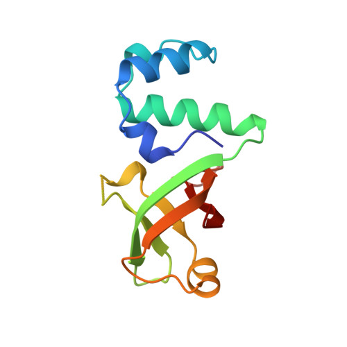
    The E. coli Rho protein disengages newly transcribed RNA from its DNA template, helping terminate certain transcripts. We have determined the X-ray crystal structure of the RNA-binding domain of Rho complexed to an RNA ligand. Filters that screen both ligand size and chemical functionality line the primary nucleic acid-binding site, imparting sequence specificity to a generic single-stranded nucleic acid-binding fold and explaining the preference of Rho for cytosine-rich RNA. The crystal packing reveals two Rho domain protomers bound to a single RNA with a single base spacer, suggesting that the strong RNA-binding sites of Rho may arise from pairing of RNA-binding modules. Dimerization of symmetric subunits on an asymmetric ligand is developed as a model for allosteric control in the action of the intact Rho hexamer.


  • Organizational Affiliation
    • Whitehead Institute for Biomedical Research, 9 Cambridge Center, Massachusetts 02142, USA.

Macromolecule Content 

  • Total Structure Weight: 27.22 kDa 
  • Atom Count: 1,902 
  • Modeled Residue Count: 231 
  • Deposited Residue Count: 242 
  • Unique protein chains: 1

Macromolecules

Find similar proteins by:|  3D Structure
Entity ID: 1
MoleculeChains  Sequence LengthOrganismDetailsImage
TRANSCRIPTION TERMINATION FACTOR RHO
A, B
121Escherichia coliMutation(s): 0 
EC: 3.6.4
UniProt
Find proteins for P0AG30 (Escherichia coli (strain K12))
Explore P0AG30 
Go to UniProtKB:  P0AG30
Entity Groups
Sequence Clusters30% Identity50% Identity70% Identity90% Identity95% Identity100% Identity
UniProt GroupP0AG30
Sequence Annotations
Expand
Reference Sequence

Experimental Data & Validation

Experimental Data

  • Method: X-RAY DIFFRACTION
  • Resolution: 2.00 Å
  • R-Value Free:  0.261 (Depositor), 0.290 (DCC) 
  • R-Value Work:  0.193 (Depositor) 
Space Group: P 1
Unit Cell:
Length ( Å )Angle ( ˚ )
a = 30.42α = 99.06
b = 36.16β = 102.64
c = 54.25γ = 96.43
Software Package:
Software NamePurpose
CCP4model building
REFMACrefinement
DENZOdata reduction
SCALEPACKdata scaling
CCP4phasing

Structure Validation

View Full Validation Report


Entry History 

Deposition Data

Revision History  (Full details and data files)

  • Version 1.0: 1999-05-04
    Type: Initial release
  • Version 1.1: 2008-03-24
    Changes: Version format compliance
  • Version 1.2: 2011-07-13
    Changes: Version format compliance
  • Version 1.3: 2024-02-07
    Changes: Data collection, Database references, Derived calculations, Other