1A73

INTRON-ENCODED ENDONUCLEASE I-PPOI COMPLEXED WITH DNA


Experimental Data Snapshot

  • Method: X-RAY DIFFRACTION
  • Resolution: 1.80 Å
  • R-Value Free: 0.297 
  • R-Value Work: 0.208 
  • R-Value Observed: 0.208 

wwPDB Validation   3D Report Full Report


This is version 1.3 of the entry. See complete history


Literature

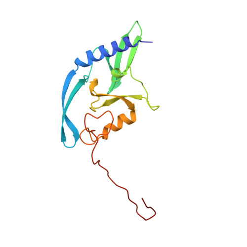
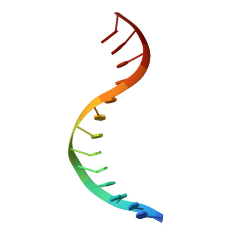
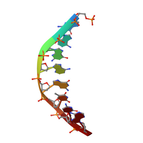
DNA binding and cleavage by the nuclear intron-encoded homing endonuclease I-PpoI.

Flick, K.E.Jurica, M.S.Monnat Jr., R.J.Stoddard, B.L.

(1998) Nature 394: 96-101

  • DOI: https://doi.org/10.1038/27952
  • Primary Citation of Related Structures:  
    1A73, 1A74, 1IPP

  • PubMed Abstract: 

    Homing endonucleases are a diverse collection of proteins that are encoded by genes with mobile, self-splicing introns. They have also been identified in self-splicing inteins (protein introns). These enzymes promote the movement of the DNA sequences that encode them from one chromosome location to another; they do this by making a site-specific double-strand break at a target site in an allele that lacks the corresponding mobile intron. The target sites recognized by these small endonucleases are generally long (14-44 base pairs). Four families of homing endonucleases have been identified, including the LAGLIDADG, the His-Cys box, the GIY-YIG and the H-N-H endonucleases. The first identified His-Cys box homing endonuclease was I-PpoI from the slime mould Physarum polycephalum. Its gene resides in one of only a few nuclear introns known to exhibit genetic mobility. Here we report the structure of the I-PpoI homing endonuclease bound to homing-site DNA determined to 1.8 A resolution. I-PpoI displays an elongated fold of dimensions 25 x 35 x 80 A, with mixed alpha/beta topology. Each I-PpoI monomer contains three antiparallel beta-sheets flanked by two long alpha-helices and a long carboxy-terminal tail, and is stabilized by two bound zinc ions 15 A apart. The enzyme possesses a new zinc-bound fold and endonuclease active site. The structure has been determined in both uncleaved substrate and cleaved product complexes.


  • Organizational Affiliation

    Fred Hutchinson Cancer Research Center, University of Washington, Seattle 98109, USA.


Macromolecules

Find similar proteins by:  (by identity cutoff)  |  3D Structure
Entity ID: 3
MoleculeChains Sequence LengthOrganismDetailsImage
INTRON 3 (I-PPO) ENCODED ENDONUCLEASEE [auth A],
F [auth B]
163Physarum polycephalumMutation(s): 0 
UniProt
Find proteins for Q94702 (Physarum polycephalum)
Explore Q94702 
Go to UniProtKB:  Q94702
Entity Groups  
Sequence Clusters30% Identity50% Identity70% Identity90% Identity95% Identity100% Identity
UniProt GroupQ94702
Sequence Annotations
Expand
  • Reference Sequence

Find similar nucleic acids by:  Sequence   |   3D Structure  

Entity ID: 1
MoleculeChains LengthOrganismImage
DNA (5'-D(*TP*TP*GP*AP*CP*TP*CP*TP*CP*TP*TP*AP*A)-3')A [auth C],
C [auth E]
13N/A
Sequence Annotations
Expand
  • Reference Sequence

Find similar nucleic acids by:  Sequence   |   3D Structure  

Entity ID: 2
MoleculeChains LengthOrganismImage
DNA (5'-D(P*GP*AP*GP*AP*GP*TP*CP*A)-3')B [auth D],
D [auth F]
8N/A
Sequence Annotations
Expand
  • Reference Sequence
Experimental Data & Validation

Experimental Data

  • Method: X-RAY DIFFRACTION
  • Resolution: 1.80 Å
  • R-Value Free: 0.297 
  • R-Value Work: 0.208 
  • R-Value Observed: 0.208 
  • Space Group: P 31 2 1
Unit Cell:
Length ( Å )Angle ( ˚ )
a = 113.7α = 90
b = 113.7β = 90
c = 88.4γ = 120
Software Package:
Software NamePurpose
CCP4model building
X-PLORrefinement
MOSFLMdata reduction
CCP4data scaling
ROTAVATAdata scaling
CCP4phasing

Structure Validation

View Full Validation Report


Entry History 

Deposition Data

Revision History  (Full details and data files)

  • Version 1.0: 1998-10-14
    Type: Initial release
  • Version 1.1: 2008-05-22
    Changes: Version format compliance
  • Version 1.2: 2011-07-13
    Changes: Version format compliance
  • Version 1.3: 2024-02-07
    Changes: Data collection, Database references, Derived calculations, Refinement description