Ribonuclease P protein structure: evolutionary origins in the translational apparatus.
Stams, T., Niranjanakumari, S., Fierke, C.A., Christianson, D.W.(1998) Science 280: 752-755
- PubMed: 9563955 Search on PubMed
- DOI: https://doi.org/10.1126/science.280.5364.752
- Primary Citation Related Structures:
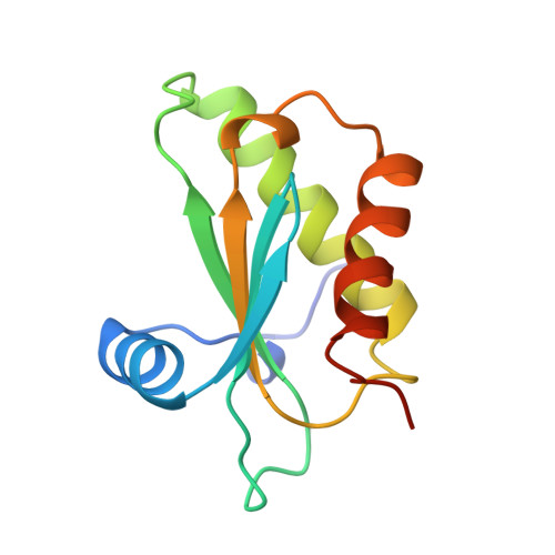
1A6F - PubMed Abstract:
The crystal structure of Bacillus subtilis ribonuclease P protein is reported at 2.6 angstroms resolution. This protein binds to ribonuclease P RNA to form a ribonucleoprotein holoenzyme with optimal catalytic activity. Mutagenesis and biochemical data indicate that an unusual left-handed betaalphabeta crossover connection and a large central cleft in the protein form conserved RNA binding sites; a metal binding loop may comprise a third RNA binding site. The unusual topology is partly shared with ribosomal protein S5 and the ribosomal translocase elongation factor G, which suggests evolution from a common RNA binding ancestor in the primordial translational apparatus.
- Roy and Diana Vagelos Laboratories, Department of Chemistry, University of Pennsylvania, Philadelphia, PA 19104-6323, USA.
Organizational Affiliation: