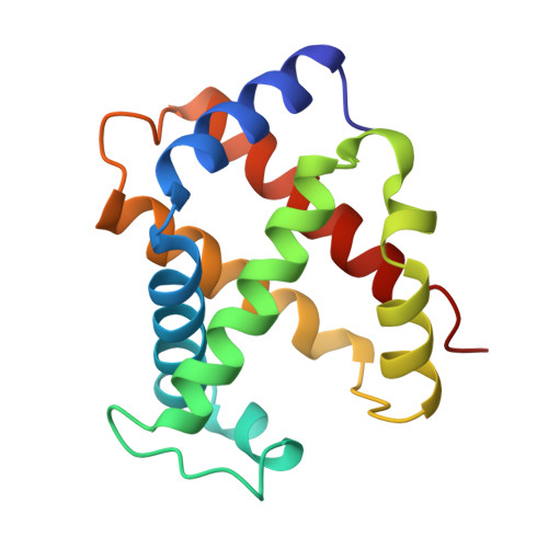
1A0U

HEMOGLOBIN (VAL BETA1 MET) MUTANT


Experimental Data Snapshot

  • Method: X-RAY DIFFRACTION
  • Resolution: 2.14 Å
  • R-Value Free: 0.239 
  • R-Value Work: 0.172 
  • R-Value Observed: 0.179 

Starting Model: experimental
View more details

wwPDB Validation   3D Report Full Report


Ligand Structure Quality Assessment 


This is version 1.4 of the entry. See complete history


Literature

High-resolution crystal structures of human hemoglobin with mutations at tryptophan 37beta: structural basis for a high-affinity T-state,.

Kavanaugh, J.S.Weydert, J.A.Rogers, P.H.Arnone, A.

(1998) Biochemistry 37: 4358-4373

  • DOI: https://doi.org/10.1021/bi9708702
  • Primary Citation of Related Structures:  
    1A00, 1A01, 1A0U, 1A0Z

  • PubMed Abstract: 

    The high-resolution X-ray structures of the deoxy forms of four recombinant hemoglobins in which Trp37(C3)beta is replaced with Tyr (betaW37Y), Ala (betaW37A), Glu (betaW37E), or Gly (betaW37G) have been refined and analyzed with superposition methods that partition mutation-induced perturbations into quaternary structure changes and tertiary structure changes. In addition, a new cross-validation statistic that is sensitive to local changes in structure (a "local Rfree" parameter) was used as an objective measure of the significance of the tertiary structure changes. No significant mutation-induced changes in tertiary structure are detected at the mutation site itself for any of the four mutants studied. Instead, disruption of the intersubunit contacts associated with Trp37(C3)beta results in (1) a change in quaternary structure at the alpha1beta2 interface, (2) alpha subunit tertiary structure changes that are centered at Asp94(G1)alpha-Pro95(G2)alpha, (3) beta subunit tertiary structure changes that are located between residues Asp99(G1)beta and Asn102(G4)beta, (4) increased mobility of the alpha subunit COOH-terminal dipeptide, and (5) shortening of the Fe-Nepsilon2His(F8) bond in the alpha and beta subunits of the betaW37G and betaW37E mutants. In each case, the magnitude of the change in a particular structural parameter increases in the order betaW37Y < betaW37A < betaW37E approximately betaW37G, which corresponds closely to the degree of functional disruption documented in the preceding papers.


  • Organizational Affiliation

    Department of Biochemistry, College of Medicine, The University of Iowa, Iowa City, Iowa 52242, USA.


Macromolecules
Find similar proteins by:  (by identity cutoff)  |  3D Structure
Entity ID: 1
MoleculeChains Sequence LengthOrganismDetailsImage
HEMOGLOBIN (ALPHA CHAIN)
A, C
141Homo sapiensMutation(s): 0 
Gene Names: HUMAN BETA GLOBIN
UniProt & NIH Common Fund Data Resources
Find proteins for P69905 (Homo sapiens)
Explore P69905 
Go to UniProtKB:  P69905
PHAROS:  P69905
Entity Groups  
Sequence Clusters30% Identity50% Identity70% Identity90% Identity95% Identity100% Identity
UniProt GroupP69905
Sequence Annotations
Expand
  • Reference Sequence
Find similar proteins by:  (by identity cutoff)  |  3D Structure
Entity ID: 2
MoleculeChains Sequence LengthOrganismDetailsImage
HEMOGLOBIN (BETA CHAIN)
B, D
146Homo sapiensMutation(s): 0 
Gene Names: HUMAN BETA GLOBIN
UniProt & NIH Common Fund Data Resources
Find proteins for P68871 (Homo sapiens)
Explore P68871 
Go to UniProtKB:  P68871
PHAROS:  P68871
GTEx:  ENSG00000244734 
Entity Groups  
Sequence Clusters30% Identity50% Identity70% Identity90% Identity95% Identity100% Identity
UniProt GroupP68871
Sequence Annotations
Expand
  • Reference Sequence
Experimental Data & Validation

Experimental Data

  • Method: X-RAY DIFFRACTION
  • Resolution: 2.14 Å
  • R-Value Free: 0.239 
  • R-Value Work: 0.172 
  • R-Value Observed: 0.179 
  • Space Group: P 21 21 2
Unit Cell:
Length ( Å )Angle ( ˚ )
a = 97.2α = 90
b = 99.3β = 90
c = 65.9γ = 90
Software Package:
Software NamePurpose
PROLSQrefinement
SDMSdata reduction
SDMSdata scaling

Structure Validation

View Full Validation Report


Ligand Structure Quality Assessment 


Entry History 

Deposition Data

Revision History  (Full details and data files)

  • Version 1.0: 1998-03-18
    Type: Initial release
  • Version 1.1: 2008-03-24
    Changes: Version format compliance
  • Version 1.2: 2011-07-13
    Changes: Version format compliance
  • Version 1.3: 2023-08-02
    Changes: Database references, Derived calculations, Refinement description
  • Version 1.4: 2024-05-22
    Changes: Data collection