158D | pdb_0000158d

CRYSTALLOGRAPHIC ANALYSIS OF C-C-A-A-G-C-T-T-G-G AND ITS IMPLICATIONS FOR BENDING IN B-DNA


Experimental Data Snapshot

  • Method: X-RAY DIFFRACTION
  • Resolution: 1.90 Å
  • R-Value Work: 
    0.170 (DCC) 
  • R-Value Observed: 
    0.179 (Depositor) 

wwPDB Validation 3D Report Full Report

Validation slider image for 158D

This is version 1.3 of the entry. See complete history

Literature

Crystallographic analysis of C-C-A-A-G-C-T-T-G-G and its implications for bending in B-DNA.

Grzeskowiak, K.Goodsell, D.S.Kaczor-Grzeskowiak, M.Cascio, D.Dickerson, R.E.

(1993) Biochemistry 32: 8923-8931

  • DOI: https://doi.org/10.1021/bi00085a025
  • Primary Citation Related Structures: 
    158D

  • PubMed Abstract: 

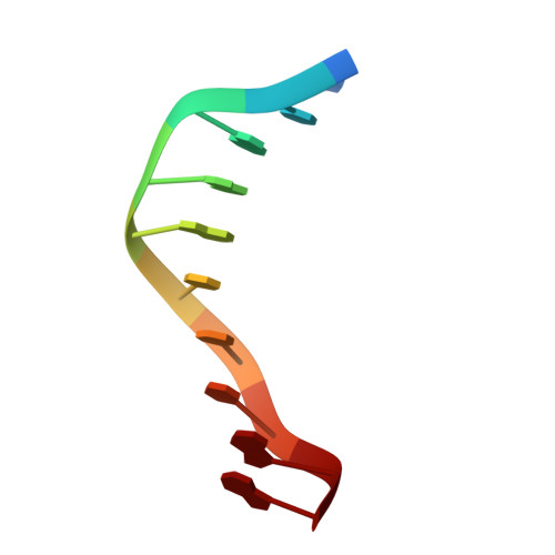
    Stacked B-DNA double helices of sequence C-C-A-A-G-C-T-T-G-G exhibit the same 23 degrees bend at -T-G-G C-C-A- across the nonbonded junction between helices that is observed in the middle of the decamer helix of sequence C-A-T-G-G-C-C-A-T-G, even though the space group (hexagonal vs orthorhombic), crystal packing, and connectedness at the center of the bent segment are quite different. An identical bend occurs across the interhelix junction of every monoclinic crystal structure of sequence C-C-A-x-x-x-x-T-G-G, suggesting that T-G-G-C-C-A constitutes a natural bending element in B-DNA. The bend occurs by rolling stacked base pairs about their long axes; there is no "tilt" component. Of the three possible models for A-tract bending--bent-A-tract, junction bends, or bent-non-A--which cannot be distinguished by solution measurements, all crystallographic evidence over the past 10 years unanimously supports the non-A regions as the actual bending loci.


  • Organizational Affiliation
    • Department of Chemistry and Biochemistry, University of California, Los Angeles 90024.

Macromolecule Content 

  • Total Structure Weight: 6.17 kDa 
  • Atom Count: 481 
  • Modeled Residue Count: 20 
  • Deposited Residue Count: 20 
  • Unique nucleic acid chains: 1

Macromolecules

Find similar nucleic acids by:  Sequence
Entity ID: 1
MoleculeChains LengthOrganismImage
DNA (5'-D(*CP*CP*AP*AP*GP*CP*TP*TP*GP*G)-3')
A, B
10N/A
Sequence Annotations
Expand
Reference Sequence

Experimental Data & Validation

Experimental Data

  • Method: X-RAY DIFFRACTION
  • Resolution: 1.90 Å
  • R-Value Work:  0.170 (DCC) 
  • R-Value Observed: 0.179 (Depositor) 
Space Group: P 6
Unit Cell:
Length ( Å )Angle ( ˚ )
a = 53.08α = 90
b = 53.08β = 90
c = 34.32γ = 120
Software Package:
Software NamePurpose
NUCLSQrefinement

Structure Validation

View Full Validation Report


Entry History 

Deposition Data

Revision History  (Full details and data files)

  • Version 1.0: 1994-05-31
    Type: Initial release
  • Version 1.1: 2008-05-22
    Changes: Version format compliance
  • Version 1.2: 2011-07-13
    Changes: Version format compliance
  • Version 1.3: 2024-02-07
    Changes: Data collection, Database references, Derived calculations