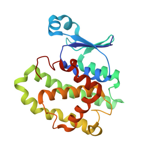
13GS

GLUTATHIONE S-TRANSFERASE COMPLEXED WITH SULFASALAZINE


Experimental Data Snapshot

  • Method: X-RAY DIFFRACTION
  • Resolution: 1.90 Å
  • R-Value Free: 0.222 
  • R-Value Work: 0.193 
  • R-Value Observed: 0.193 

Starting Model: experimental
View more details

wwPDB Validation   3D Report Full Report


Ligand Structure Quality Assessment 


This is version 1.5 of the entry. See complete history


Literature

The Ligandin (Non-Substrate) Binding Site of Human Pi Class Glutathione Transferase is Located in the Electrophile Binding Site (H-Site).

Oakley, A.J.Lo Bello, M.Nuccetelli, M.Mazzetti, A.P.Parker, M.W.

(1999) J Mol Biol 291: 913

  • DOI: https://doi.org/10.1006/jmbi.1999.3029
  • Primary Citation of Related Structures:  
    12GS, 13GS, 18GS, 19GS, 20GS

  • PubMed Abstract: 

    Glutathione S -transferases (GSTs) play a pivotal role in the detoxification of foreign chemicals and toxic metabolites. They were originally termed ligandins because of their ability to bind large molecules (molecular masses >400 Da), possibly for storage and transport roles. The location of the ligandin site in mammalian GSTs is still uncertain despite numerous studies in recent years. Here we show by X-ray crystallography that the ligandin binding site in human pi class GST P1-1 occupies part of one of the substrate binding sites. This work has been extended to the determination of a number of enzyme complex crystal structures which show that very large ligands are readily accommodated into this substrate binding site and in all, but one case, causes no significant movement of protein side-chains. Some of these molecules make use of a hitherto undescribed binding site located in a surface pocket of the enzyme. This site is conserved in most, but not all, classes of GSTs suggesting it may play an important functional role.


  • Organizational Affiliation

    The Ian Potter Foundation Protein Crystallography Laboratory, St. Vincent's Institute of Medical Research, Fitzroy, Victoria, 3065, Australia.


Macromolecules
Find similar proteins by:  (by identity cutoff)  |  3D Structure
Entity ID: 1
MoleculeChains Sequence LengthOrganismDetailsImage
GLUTATHIONE S-TRANSFERASE
A, B
210Homo sapiensMutation(s): 0 
Gene Names: GSTP1
EC: 2.5.1.18
UniProt & NIH Common Fund Data Resources
Find proteins for P09211 (Homo sapiens)
Explore P09211 
Go to UniProtKB:  P09211
PHAROS:  P09211
GTEx:  ENSG00000084207 
Entity Groups  
Sequence Clusters30% Identity50% Identity70% Identity90% Identity95% Identity100% Identity
UniProt GroupP09211
Sequence Annotations
Expand
  • Reference Sequence
Small Molecules
Binding Affinity Annotations 
IDSourceBinding Affinity
SAS PDBBind:  13GS Ki: 2.40e+4 (nM) from 1 assay(s)
Experimental Data & Validation

Experimental Data

  • Method: X-RAY DIFFRACTION
  • Resolution: 1.90 Å
  • R-Value Free: 0.222 
  • R-Value Work: 0.193 
  • R-Value Observed: 0.193 
  • Space Group: C 1 2 1
Unit Cell:
Length ( Å )Angle ( ˚ )
a = 79.455α = 90
b = 91.107β = 98.44
c = 69.559γ = 90
Software Package:
Software NamePurpose
X-PLORmodel building
X-PLORrefinement
DENZOdata reduction
SCALEPACKdata scaling
X-PLORphasing

Structure Validation

View Full Validation Report


Ligand Structure Quality Assessment 


Entry History 

Deposition Data

Revision History  (Full details and data files)

  • Version 1.0: 1999-01-13
    Type: Initial release
  • Version 1.1: 2008-03-03
    Changes: Version format compliance
  • Version 1.2: 2011-07-13
    Changes: Database references, Derived calculations, Non-polymer description, Version format compliance
  • Version 1.3: 2011-12-21
    Changes: Non-polymer description, Structure summary
  • Version 1.4: 2023-08-02
    Changes: Database references, Derived calculations, Other, Refinement description
  • Version 1.5: 2024-05-22
    Changes: Data collection