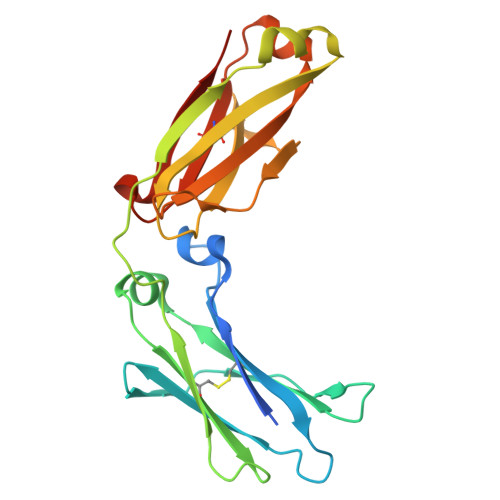
13AN | pdb_000013an

Crystal Structure of Human IGG1 FC Fragment (P33A, Orthorhombic C Form)


Experimental Data Snapshot

  • Method: X-RAY DIFFRACTION
  • Resolution: 2.35 Å
  • R-Value Free: 
    0.277 (Depositor), 0.276 (DCC) 
  • R-Value Work: 
    0.218 (Depositor), 0.221 (DCC) 
  • R-Value Observed: 
    0.221 (Depositor) 

Starting Model: experimental
View more details

wwPDB Validation 3D Report Full Report

Validation slider image for 13AN

This is version 1.0 of the entry. See complete history. 

Literature

Near UV Photo-Degradation of IgG1-Fc Promoted by His and Fe(III): Probing the Role of the Metal-Binding Pocket by Site-Directed Mutagenesis.

Ballesteros, M.E., Rodriguez, M.S., Seibold, S., Battaile, K.P., Lovell, S., Tolbert, T.J., Schoneich, C.

(2026) Mol Pharm 23: 5489-5501

  • DOI: https://doi.org/10.1021/acs.molpharmaceut.6c00605
  • Primary Citation Related Structures: 
    13AL, 13AM, 13AN

  • PubMed Abstract: 

    Light exposure of therapeutic proteins can affect chemical and physical stability and compromise safety and efficacy. In order to develop stable pharmaceutical formulations without undesired side effects, it is essential to understand both product formation and reaction mechanisms which lead to light-induced protein degradation. We recently reported that visible and near UV light induce site-specific fragmentation and photo-conversion of a heavy chain Thr residue in IgG1-Fc, full length IgG1 and IgG1-based fusion proteins formulated in Fe(III)-containing histidine buffer. These post-translational modifications manifest differently in each modality, with the photo-conversion of Thr to Ala being more prominent in IgG1-Fc. Mechanistically, these photo-degradation pathways occur within a Fe(III)-binding pocket present in the CH2 domain. Here, we investigate the role of individual metal-binding residues located in the Fe(III)-binding pocket in the photo-degradation of IgG1-Fc. The single mutants of Asp25 to Ala, Thr32 to Ala, and Glu34 to Gln in IgG1-Fc completely prevent the site-specific photo-degradation of Thr32, while the mutation of Pro33 to Ala had only a relatively small effect. A comparison of crystal structures reveals that the replacement of Pro33 by Ala does not significantly affect the structure of the Fe(III)-binding pocket. Our results indicate that a single amino acid mutation can prevent the Fe(III)-catalyzed photo-degradation, suggesting that single point mutations may be a future pathway towards photo-stable pharmaceutical formulations.


  • Organizational Affiliation: 
    • Department of Pharmaceutical Chemistry, University of Kansas, 2093 Constant Avenue, Lawrence, Kansas66047, United States.

Macromolecule Content 

  • Total Structure Weight: 52.57 kDa 
  • Atom Count: 3,546 
  • Modeled Residue Count: 414 
  • Deposited Residue Count: 446 
  • Unique protein chains: 1

Macromolecules

Find similar proteins by:|  3D Structure
Entity ID: 1
MoleculeChains  Sequence LengthOrganismDetailsImage
Hepatitis B virus receptor binding protein
A, B
223Homo sapiensMutation(s): 1 
UniProt
Find proteins for Q6PYX1 (Homo sapiens)
Explore Q6PYX1 
Go to UniProtKB:  Q6PYX1
Entity Groups
Sequence Clusters30% Identity50% Identity70% Identity90% Identity95% Identity100% Identity
UniProt GroupQ6PYX1
Glycosylation
Glycosylation Sites: 1
Sequence Annotations
Expand
Reference Sequence

Oligosaccharides

Help  
Entity ID: 2
MoleculeChains Length2D Diagram GlycosylationD Interactions
2-acetamido-2-deoxy-beta-D-glucopyranose-(1-2)-alpha-D-mannopyranose-(1-3)-[2-acetamido-2-deoxy-beta-D-glucopyranose-(1-2)-alpha-D-mannopyranose-(1-6)]beta-D-mannopyranose-(1-4)-2-acetamido-2-deoxy-beta-D-glucopyranose-(1-4)-2-acetamido-2-deoxy-beta-D-glucopyranose
C
7N-Glycosylation
Entity ID: 3
MoleculeChains Length2D Diagram GlycosylationD Interactions
2-acetamido-2-deoxy-beta-D-glucopyranose-(1-2)-alpha-D-mannopyranose-(1-3)-[alpha-D-mannopyranose-(1-6)]beta-D-mannopyranose-(1-4)-2-acetamido-2-deoxy-beta-D-glucopyranose-(1-4)-2-acetamido-2-deoxy-beta-D-glucopyranose
D
6N-Glycosylation

Experimental Data & Validation

Experimental Data

  • Method: X-RAY DIFFRACTION
  • Resolution: 2.35 Å
  • R-Value Free:  0.277 (Depositor), 0.276 (DCC) 
  • R-Value Work:  0.218 (Depositor), 0.221 (DCC) 
  • R-Value Observed: 0.221 (Depositor) 
Space Group: C 2 2 21
Unit Cell:
Length ( Å )Angle ( ˚ )
a = 64.437α = 90
b = 113.94β = 90
c = 158.097γ = 90
Software Package:
Software NamePurpose
PHENIXrefinement
Aimlessdata scaling
XDSdata reduction
PHASERphasing
PDB_EXTRACTdata extraction

Structure Validation

View Full Validation Report


Entry History 

& Funding Information

Deposition Data


Funding OrganizationLocationGrant Number
National Institutes of Health/Office of the DirectorUnited StatesS10OD030394

Revision History  (Full details and data files)

  • Version 1.0: 2026-11-11
    Type: Initial release