121D | pdb_0000121d

MOLECULAR STRUCTURE OF THE A-TRACT DNA DODECAMER D(CGCAAATTTGCG) COMPLEXED WITH THE MINOR GROOVE BINDING DRUG NETROPSIN


Experimental Data Snapshot

  • Method: X-RAY DIFFRACTION
  • Resolution: 2.20 Å
  • R-Value Work: 
    0.198 (Depositor) 
  • R-Value Observed: 
    0.198 (Depositor) 

wwPDB Validation 3D Report Full Report

Validation slider image for 121D

This is version 1.4 of the entry. See complete history. 

Literature

Molecular structure of the A-tract DNA dodecamer d(CGCAAATTTGCG) complexed with the minor groove binding drug netropsin.

Tabernero, L., Verdaguer, N., Coll, M., Fita, I., van der Marel, G.A., van Boom, J.H., Rich, A., Aymami, J.

(1993) Biochemistry 32: 8403-8410

  • DOI: https://doi.org/10.1021/bi00084a004
  • Primary Citation Related Structures: 
    121D

  • PubMed Abstract: 

    The molecular structure of the complex between the minor groove binding drug netropsin and the dodecamer d(CGCAAATTTGCG) has been solved and refined by X-ray diffraction analysis to an R-factor of 19.8% and 2.2-A resolution. The drug lies in the narrow minor groove of the B-DNA fragment, covering five of the six A.T base pairs (from A5.T20 to T9.A16). The long six A.T base pair tract allows the drug to bind in a position that optimizes its contacts with the DNA, establishing hydrogen bonds with O2 of thymines and N3 of adenines. The DNA molecule shows a high propeller twist only at the A6.T19 step of the A-tract. Two three-centered hydrogen bonds are observed in the major groove at half of the A-tract.


  • Organizational Affiliation: 
    • Departament d'Enginyeria Química, Universitat Politècnica de Catalunya, Barcelona, Spain.

Macromolecule Content 

  • Total Structure Weight: 7.76 kDa 
  • Atom Count: 564 
  • Modeled Residue Count: 24 
  • Deposited Residue Count: 24 
  • Unique nucleic acid chains: 1

Macromolecules

Find similar nucleic acids by:  Sequence
Entity ID: 1
MoleculeChains LengthOrganismImage
DNA (5'-D(*CP*GP*CP*AP*AP*AP*TP*TP*TP*GP*CP*G)-3')
A, B
12N/A
Sequence Annotations
Expand
Reference Sequence

Small Molecules

Ligands 1 Unique
IDChains Name / Formula / InChI Key2D Diagram3D Interactions
NT

Query on NT


Download:Ideal Coordinates CCD File
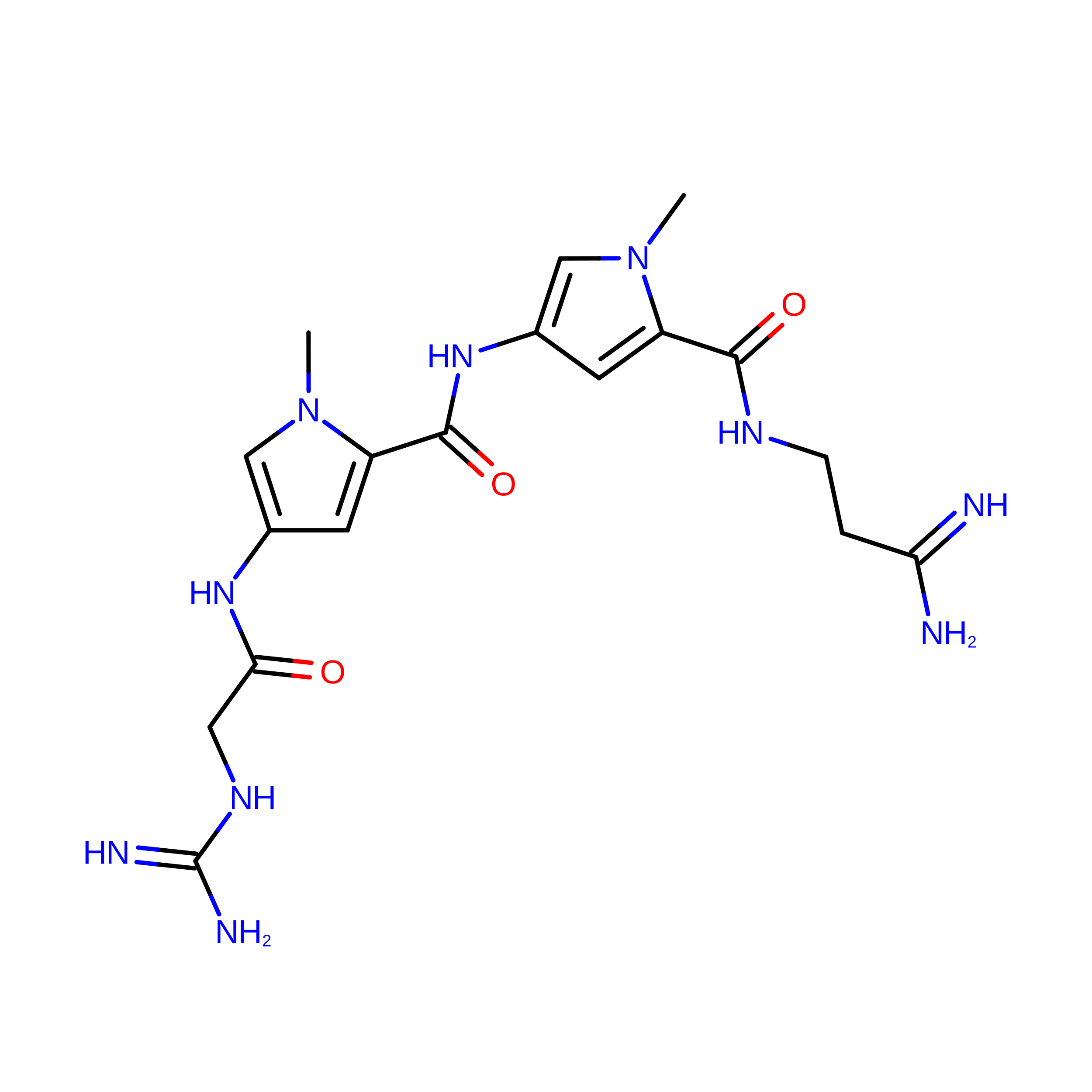
C [auth A]NETROPSIN
C18 H26 N10 O3
IDBIFFKSXLYUOT-UHFFFAOYSA-N

Experimental Data & Validation

Experimental Data

  • Method: X-RAY DIFFRACTION
  • Resolution: 2.20 Å
  • R-Value Work:  0.198 (Depositor) 
  • R-Value Observed: 0.198 (Depositor) 
Space Group: P 21 21 21
Unit Cell:
Length ( Å )Angle ( ˚ )
a = 25.65α = 90
b = 42.03β = 90
c = 65.33γ = 90
Software Package:
Software NamePurpose
X-PLORrefinement

Structure Validation

View Full Validation Report


Entry History 

Deposition Data

Revision History  (Full details and data files)

  • Version 1.0: 1994-01-15
    Type: Initial release
  • Version 1.1: 2008-05-22
    Changes: Version format compliance
  • Version 1.2: 2011-07-13
    Changes: Version format compliance
  • Version 1.3: 2018-04-18
    Changes: Advisory, Data collection
  • Version 1.4: 2024-02-07
    Changes: Advisory, Data collection, Database references, Derived calculations