11UF | pdb_000011uf

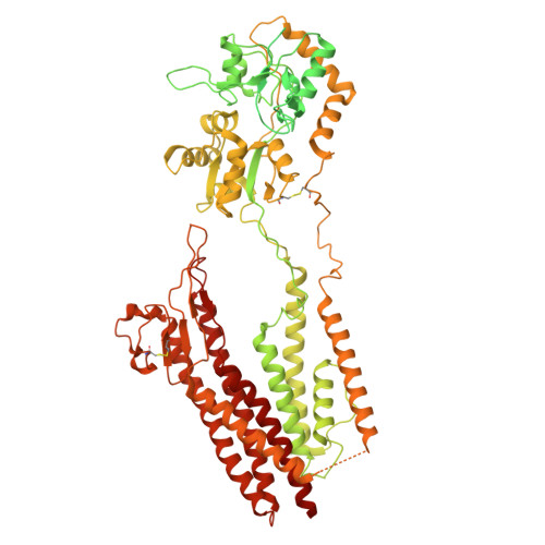
GluA2-y5-CNIH1 open state + RR2b + Glu (LBD-TMD)

  • Classification: MEMBRANE PROTEIN
  • Organism(s): Rattus norvegicus, Homo sapiens
  • Expression System: Homo sapiens
  • Mutation(s): No 

  • Deposited: 2026-03-13 Released: 2026-09-23 
  • Deposition Author(s): Gangwar, S.P., Newton, T.P., Yen, L.Y., Sobolevsky, A.I.
  • Funding Organization(s): National Institutes of Health/National Institute of Neurological Disorders and Stroke (NIH/NINDS), National Institutes of Health/National Institute of Arthritis and Musculoskeletal and Skin Diseases (NIH/NIAMS), National Institutes of Health/National Cancer Institute (NIH/NCI)

Experimental Data Snapshot

  • Method: ELECTRON MICROSCOPY
  • Resolution: 3.39 Å
  • Aggregation State: PARTICLE 
  • Reconstruction Method: SINGLE PARTICLE 

wwPDB Validation 3D Report Full Report

Validation slider image for 11UF

This is version 1.1 of the entry. See complete history. 

Literature

Tuning of AMPA receptor activation by inhibitory auxiliary subunits and epilepsy-associated disease mutations.

Newton, T.P., Yelshanskaya, M.V., Aktolun, M., Gangwar, S.P., Yen, L.Y., Alekseev, A.A., Sobolevsky, I.A., Kurnikova, M.G., Sobolevsky, A.I.

(2026) Nat Commun 17

  • DOI: https://doi.org/10.1038/s41467-026-77416-0
  • Primary Citation Related Structures: 
    11UF, 11UG, 11UH, 11UI

  • PubMed Abstract: 

    AMPA receptors (AMPARs) facilitate excitatory neurotransmission and are implicated in neurological and neuropsychiatric disorders. Binding of the neurotransmitter glutamate triggers AMPAR activation, which is modulated by potentiating and inhibitory auxiliary subunits. While modulation by potentiating auxiliary subunits has been studied extensively, activation involving inhibitory subunits has not been examined. Here, we present structures of the activated GluA2 AMPAR alone and in complex with inhibitory subunits GSG1L or γ5. Our results suggest that gating kinetics strongly depend on auxiliary subunit identity, while pore size and conductance do not. We find that receptor deactivation becomes slower if an auxiliary subunit promotes tightening of the interface between two ligand-binding domain dimers during activation. In addition, we functionally characterize the effects of epilepsy-associated disease mutations on AMPAR activation and desensitization kinetics, displaying strong dependence on the type of bound auxiliary subunit. Modulation by auxiliary subunits therefore has to be considered when designing AMPAR-targeting therapeutics.


  • Organizational Affiliation: 
    • Department of Biochemistry and Molecular Biophysics, Columbia University, New York, NY, USA.

Macromolecule Content 

  • Total Structure Weight: 504.93 kDa 
  • Atom Count: 18,310 
  • Modeled Residue Count: 2,318 
  • Deposited Residue Count: 4,484 
  • Unique protein chains: 2

Macromolecules

Find similar proteins by:|  3D Structure
Entity ID: 1
MoleculeChains  Sequence LengthOrganismDetailsImage
Isoform Flip of Glutamate receptor 2,Voltage-dependent calcium channel gamma-5 subunit
A, B, C, D
1,049Rattus norvegicusMutation(s): 0 
Gene Names: Gria2, GluA2, Glur2, Cacng5
UniProt & NIH Common Fund Data Resources
Find proteins for P19491 (Rattus norvegicus)
Explore P19491 
Go to UniProtKB:  P19491
Find proteins for Q8VHW4 (Mus musculus)
Explore Q8VHW4 
Go to UniProtKB:  Q8VHW4
IMPC:  MGI:2157946
Entity Groups
Sequence Clusters30% Identity50% Identity70% Identity90% Identity95% Identity100% Identity
UniProt GroupsP19491Q8VHW4
Sequence Annotations
Expand
Reference Sequence
Find similar proteins by:|  3D Structure
Entity ID: 2
MoleculeChains  Sequence LengthOrganismDetailsImage
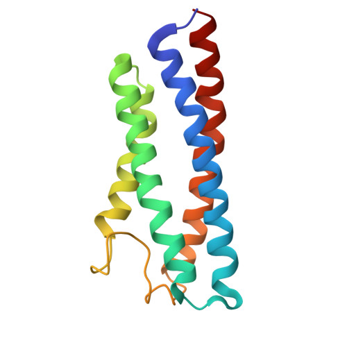
Protein cornichon homolog 1
E, F
144Homo sapiensMutation(s): 0 
UniProt & NIH Common Fund Data Resources
Find proteins for O95406 (Homo sapiens)
Explore O95406 
Go to UniProtKB:  O95406
PHAROS:  O95406
GTEx:  ENSG00000100528 
Entity Groups
Sequence Clusters30% Identity50% Identity70% Identity90% Identity95% Identity100% Identity
UniProt GroupO95406
Sequence Annotations
Expand
Reference Sequence

Experimental Data & Validation

Experimental Data

  • Method: ELECTRON MICROSCOPY
  • Resolution: 3.39 Å
  • Aggregation State: PARTICLE 
  • Reconstruction Method: SINGLE PARTICLE 
EM Software:
TaskSoftware PackageVersion
MODEL REFINEMENTPHENIX1.11.1_2575
RECONSTRUCTIONcryoSPARC

Structure Validation

View Full Validation Report


Entry History 

& Funding Information

Deposition Data


Funding OrganizationLocationGrant Number
National Institutes of Health/National Institute of Neurological Disorders and Stroke (NIH/NINDS)United StatesNS139087
National Institutes of Health/National Institute of Neurological Disorders and Stroke (NIH/NINDS)United StatesF31NS147755
National Institutes of Health/National Institute of Neurological Disorders and Stroke (NIH/NINDS)United StatesF31NS132554
National Institutes of Health/National Institute of Neurological Disorders and Stroke (NIH/NINDS)United StatesNS083660
National Institutes of Health/National Institute of Neurological Disorders and Stroke (NIH/NINDS)United StatesNS107253
National Institutes of Health/National Institute of Arthritis and Musculoskeletal and Skin Diseases (NIH/NIAMS)United StatesAR078814
National Institutes of Health/National Cancer Institute (NIH/NCI)United StatesCA206573

Revision History  (Full details and data files)

  • Version 1.0: 2026-09-23
    Type: Initial release
  • Version 1.1: 2026-10-21
    Changes: Data collection, Database references