Download Mendeley
Cofactor-Free Tau Filaments Are Dynamic and Undergo Structural Evolution Driven by Thermodynamic Control.
Powell, W.C., Yan, N., Tse, E., Melo, A.A., Vasquez, J.A., Southworth, D.R., Gestwicki, J.E.(2026) J Am Chem Soc 148: 35386-35398
- PubMed: 42677560 Search on PubMedSearch on PubMed Central
- DOI: https://doi.org/10.1021/jacs.6c04928
- Primary Citation Related Structures:
11LU, 11LV, 11LW - PubMed Abstract:
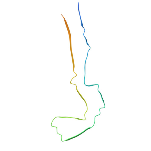
Tau filaments are a hallmark of neurodegenerative tauopathies, such as Alzheimer's disease (AD). Structural studies have revealed that patient-derived tau fibrils adopt distinct folds in different tauopathies; however, it is unclear what forces guide this process. To explore this question, we investigated the assembly of a tau fragment containing four disease-associated phospho-mimetics (termed Tau(297-407)-4D) in vitro. Under cofactor-free and quiescent conditions, Tau(297-407)-4D forms fibrils with a core structure that partially resembles the AD fold after about 7 days. Strikingly, we noticed that this filament behaves as a hydrogel and evolves into two new polymorphs as it ages over the next 35 days. Thus, tau fibrils formed under cofactor-free conditions are dynamic, exhibiting substantial nonequilibrium behavior. To probe what types of perturbations might stabilize these structures, we applied mechanical agitation, which drove the filaments toward thermodynamic equilibrium in a mechanism consistent with Ostwald ripening into solid-phase, micrometer-sized particles. Likewise, the addition of polyanionic cofactors to preformed Tau(297-407)-4D fibrils significantly stabilized them, as judged by solubility equilibria and chemical denaturation experiments. A subset of the polyanions also remodeled the fibril structure and tuned the extent of fibril-fibril interactions (i.e., "clumping"). We conclude that environmental factors, such as mechanical stress and/or polyanions, play an important role in promoting the thermodynamic stability of otherwise dynamic tau fibrils. We speculate that, in patients, such factors might contribute to the maturation of disease-specific conformers.
- Institute for Neurodegenerative Diseases, University of California San Francisco, San Francisco, California94158, United States.
Organizational Affiliation: