Download Mendeley
Emergence of neutralizing RBD antibodies following Omicron infection with limited activity against ancestral SARS-CoV-2.
Piepenbrink, M.S., Ma, Y., Panjwani, S., Blake, A.R., Bell, A.M., Kizziah, J.L., Mahmoud, S.H., Ippolito, G.C., Erdmann, N.B., Goepfert, P.A., Martinez-Sobrido, L., Kobie, J.J., Walter, M.R.(2026) iScience 29: 117166-117166
- PubMed: 42621159 Search on PubMedSearch on PubMed Central
- DOI: https://doi.org/10.1016/j.isci.2026.117166
- Primary Citation Related Structures:
11HM, 11HO, 11IS, 11IT - PubMed Abstract:
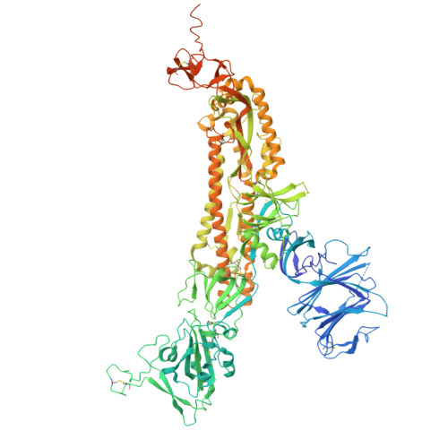
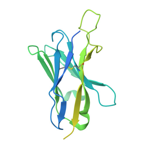
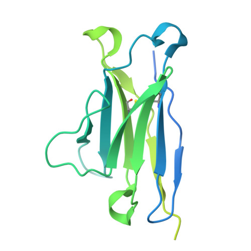
Severe acute respiratory syndrome coronavirus 2 (SARS-CoV-2) evolution reduces the efficacy of prophylactic vaccines and monoclonal antibody (mAb) therapies. To evaluate potency and breadth, receptor binding domain (RBD)-targeting neutralizing mAbs were identified from patient B cells using an Omicron KP3.1.1 Spike (S) protein bait. MAbs exhibiting the greatest neutralization potency (1332D4 and 1332E5) and binding breadth (1324A10 and 1316C10) were evaluated in greater detail. Cryo-electron microscopy (Cryo-EM) studies revealed 1332D4 and 1332E5 target RBD residue segments 439-446 and 498-506 of KP3.1.1 S, respectively. 1332E5 is a knob 498-506 -targeting class-1/4 mAb that exhibits pan-Omicron specificity but does not bind or neutralize ancestral Wuhan-1. Further analysis suggests mAbs generically defined as class 1/4 mAbs may be separated into two classes. Class 1/4 mAbs, like 1332E5, and class 4/1 mAbs that make extensive interactions with the class-4 epitope and limited contacts with the class-1 knob 498-596 . Despite these differences, the specificity of both mAb classes can be altered by mutations in knob 498-506 .
- Heersink School of Medicine, Infectious Diseases Division, University of Alabama at Birmingham, Birmingham, AL 35294, USA.
Organizational Affiliation: