Download Mendeley
Structural origins of species-specific differences in TRPV2 activation.
Fricke, T.C., Pumroy, R.A., Rocereta, J.A., De Jesus-Perez, J.J., Oprita, G., Meyer, M.J.A., Herzog, C., Echtermeyer, F.G., Hill, K., Leffler, A., Moiseenkova-Bell, V.Y.(2026) Nat Commun 17
- PubMed: 42624855 Search on PubMedSearch on PubMed Central
- DOI: https://doi.org/10.1038/s41467-026-76831-7
- Primary Citation Related Structures:
11HZ, 11IA, 11IB, 11ID, 11IE - PubMed Abstract:
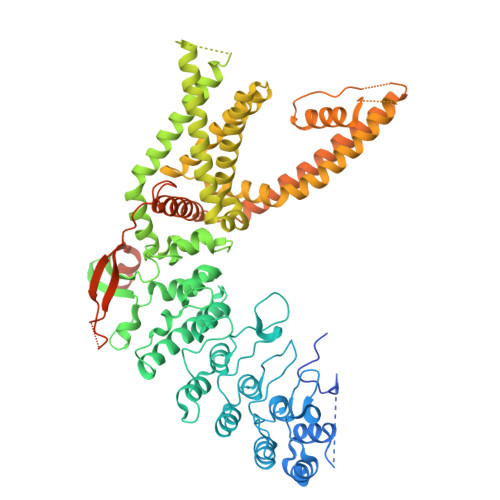
Transient receptor potential vanilloid 2 (TRPV2) is a broadly expressed ion channel implicated in diverse physiological and pathological processes. Despite strong conservation, human TRPV2 (hTRPV2) displays markedly reduced sensitivity to stimuli such as 2-aminoethoxydiphenyl borate (2-APB) and heat compared to rodent orthologs. Here we combine electrophysiology and cryo electron microscopy to define the basis of this species-dependent divergence. The structure of hTRPV2 is remarkably different at the voltage sensor-like domain (VSLD) compared to rodent channels and functional analyses show a graded activity profile between human, mouse and rat TRPV2 to a broad range of chemical and physical stimuli. Three residues located between S6 and the TRP domain tune this functional difference, as reciprocal substitutions exchange current phenotypes. hTRPV2 structures of this mutant reveal coupling between the mutation site and the VSLD as well as an additional binding site for 2-APB. Together, these findings define structural determinants of species-specific TRPV2 function.
- Department of Anesthesiology and Intensive Care Medicine, Hannover Medical School, Hannover, Germany.
Organizational Affiliation: