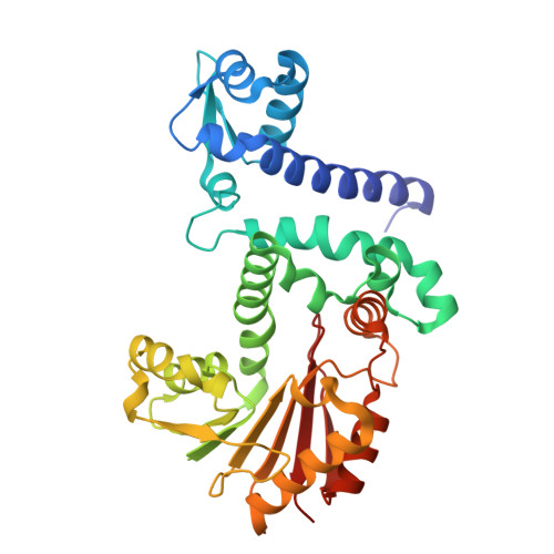
10QU | pdb_000010qu

Crystal structure of an O-methyltransferase (Dol15G) from dolastatin 15 biosynthesis in complex with S-adenosyl-L-homocysteine (SAH)


Experimental Data Snapshot

  • Method: X-RAY DIFFRACTION
  • Resolution: 2.65 Å
  • R-Value Free: 
    0.220 (Depositor), 0.219 (DCC) 
  • R-Value Work: 
    0.174 (Depositor), 0.175 (DCC) 
  • R-Value Observed: 
    0.177 (Depositor) 

Starting Model: in silico
View more details

wwPDB Validation 3D Report Full Report

Validation slider image for 10QU

Ligand Structure Quality Assessment 


This is version 1.0 of the entry. See complete history. 

Literature

Biosynthetic origin of anticancer dolastatin 15 reveals a noncanonical NRPS architecture enabling 2-keto acid activation.

Chen, M., Liu, J., Tsai, H.Y., Li, C.Y., Batucan, J.D., Eckhardt, C.W., Pham, T.H.D., Ellis, E.K., Ratnayake, R., Paul, V.J., Bruner, S.D., Donia, M.S., Luesch, H., Ding, Y.

(2026) Proc Natl Acad Sci U S A 123: e2615454123-e2615454123

  • DOI: https://doi.org/10.1073/pnas.2615454123
  • Primary Citation Related Structures: 
    10QU

  • PubMed Abstract: 

    Elucidating the biological origins and biosynthetic pathways of marine natural products remains a major challenge, particularly for compounds derived from microbial consortia. Here, we identify the marine cyanobacterial genus Dapis as the source of the potent anticancer natural product dolastatin 15 using genome-resolved metagenomics. Reconstruction of two complete and two near-complete genomes provides genome-resolved characterization of the genus Dapis , revealing extensive biosynthetic potential. Genome mining identified the dolastatin 15 biosynthetic gene cluster. Subsequent analyses reconstructed its pathway and uncovered a noncanonical five-domain nonribosomal peptide synthetase module containing an embedded 2-keto acid-activating domain, revealing an unexpected architectural solution for incorporating 2-keto acid-derived hydroxy acid building blocks. Comparative and evolutionary analyses support a model in which this architecture may have arisen through recruitment of keto acid-activating domains and reduction of canonical adenylation domain features. We further biochemically and structurally characterize an O -methyltransferase that catalyzes formation of the characteristic methoxy pyrrolinone moiety, thereby validating a key terminal tailoring step in the proposed biosynthetic pathway. Together, these findings define the biosynthetic logic of dolastatin 15 and expand understanding of substrate activation and assembly-line diversification in nonribosomal peptide biosynthesis.


  • Organizational Affiliation: 
    • Department of Medicinal Chemistry, Center for Natural Products, Drug Discovery, and Development, University of Florida, Gainesville, FL 32610.

Macromolecule Content 

  • Total Structure Weight: 75.67 kDa 
  • Atom Count: 5,594 
  • Modeled Residue Count: 667 
  • Deposited Residue Count: 676 
  • Unique protein chains: 1

Macromolecules

Find similar proteins by:|  3D Structure
Entity ID: 1
MoleculeChains  Sequence LengthOrganismDetailsImage
O-methyltransferase
A, B
338CyanobacteriotaMutation(s): 0 
Entity Groups
Sequence Clusters30% Identity50% Identity70% Identity90% Identity95% Identity100% Identity
Sequence Annotations
Expand
Reference Sequence

Small Molecules

Ligands 3 Unique
IDChains Name / Formula / InChI Key2D Diagram3D Interactions
SAH
(Subject of Investigation/LOI)

Query on SAH


Download:Ideal Coordinates CCD File
C [auth A],
H [auth B]
S-ADENOSYL-L-HOMOCYSTEINE
C14 H20 N6 O5 S
ZJUKTBDSGOFHSH-WFMPWKQPSA-N
GOL

Query on GOL


Download:Ideal Coordinates CCD File
D [auth A],
I [auth B]
GLYCEROL
C3 H8 O3
PEDCQBHIVMGVHV-UHFFFAOYSA-N
CL

Query on CL


Download:Ideal Coordinates CCD File
E [auth A],
F [auth A],
G [auth A],
J [auth B]
CHLORIDE ION
Cl
VEXZGXHMUGYJMC-UHFFFAOYSA-M

Experimental Data & Validation

Experimental Data

  • Method: X-RAY DIFFRACTION
  • Resolution: 2.65 Å
  • R-Value Free:  0.220 (Depositor), 0.219 (DCC) 
  • R-Value Work:  0.174 (Depositor), 0.175 (DCC) 
  • R-Value Observed: 0.177 (Depositor) 
Space Group: C 1 2 1
Unit Cell:
Length ( Å )Angle ( ˚ )
a = 133.015α = 90
b = 139.746β = 94.947
c = 71.145γ = 90
Software Package:
Software NamePurpose
PHENIXrefinement
REFMACrefinement
PHENIXphasing
BUCCANEERmodel building
XDSdata reduction
XDSdata scaling

Structure Validation

View Full Validation Report


Ligand Structure Quality Assessment 


Entry History 

& Funding Information

Deposition Data


Funding OrganizationLocationGrant Number
National Institutes of Health/National Institute of General Medical Sciences (NIH/NIGMS)United StatesRM1GM145426

Revision History  (Full details and data files)

  • Version 1.0: 2026-10-28
    Type: Initial release