


Crystal Structure of Apurinic endonuclease (APN1) from Babesia bovis
Lovell, S., Cooper, A., Battaile, K.P.To be published.
Experimental Data Snapshot
Starting Model: experimental
View more details
wwPDB Validation 3D Report Full Report

Macromolecule Content
Entity ID: 1 | |||||
|---|---|---|---|---|---|
| Molecule | Chains | Sequence Length | Organism | Details | Image |
| Apurinic endonuclease (APN1) | 323 | Babesia bovis T2Bo | Mutation(s): 0 Gene Names: BBOV_III005900 | 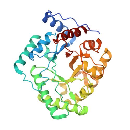 | |
UniProt | |||||
Entity Groups | |||||
| Sequence Clusters | 30% Identity50% Identity70% Identity90% Identity95% Identity100% Identity | ||||
| UniProt Group | S6B9Y3 | ||||
Sequence AnnotationsExpand | |||||
Reference Sequence | |||||
| Ligands 3 Unique | |||||
|---|---|---|---|---|---|
| ID | Chains | Name / Formula / InChI Key | 2D Diagram | 3D Interactions | |
| HEZ Download:Ideal Coordinates CCD File | DA [auth F] | HEXANE-1,6-DIOL C6 H14 O2 XXMIOPMDWAUFGU-UHFFFAOYSA-N |  | ||
| IMD Download:Ideal Coordinates CCD File | EA [auth F] J [auth A] N [auth B] R [auth C] V [auth D] | IMIDAZOLE C3 H5 N2 RAXXELZNTBOGNW-UHFFFAOYSA-O |  | ||
| ZN (Subject of Investigation/LOI) Download:Ideal Coordinates CCD File | AA [auth F] BA [auth F] CA [auth F] G [auth A] H [auth A] | ZINC ION Zn PTFCDOFLOPIGGS-UHFFFAOYSA-N |  | ||
| Length ( Å ) | Angle ( ˚ ) |
|---|---|
| a = 59.625 | α = 91.66 |
| b = 93.089 | β = 91.18 |
| c = 102.879 | γ = 91.09 |
| Software Name | Purpose |
|---|---|
| PHENIX | refinement |
| Aimless | data scaling |
| XDS | data reduction |
| PHASER | phasing |
| PDB_EXTRACT | data extraction |
| Funding Organization | Location | Grant Number |
|---|---|---|
| National Institutes of Health/National Institute Of Allergy and Infectious Diseases (NIH/NIAID) | United States | 75N93022C00036 |
| National Institutes of Health/Office of the Director | United States | S10OD030394 |


