9BQ2

Structure of the flotillin complex in a native membrane environment

  • Classification: MEMBRANE PROTEIN
  • Organism(s): Homo sapiens
  • Mutation(s): No 

  • Deposited: 2024-05-08 Released: 2024-07-24 
  • Deposition Author(s): Fu, Z., MacKinnon, R.
  • Funding Organization(s): National Institutes of Health/National Institute of General Medical Sciences (NIH/NIGMS), Howard Hughes Medical Institute (HHMI)

Experimental Data Snapshot

  • Method: ELECTRON MICROSCOPY
  • Resolution: 3.50 Å
  • Aggregation State: PARTICLE 
  • Reconstruction Method: SINGLE PARTICLE 

wwPDB Validation   3D Report Full Report


This is version 1.0 of the entry. See complete history


Literature

Structure of the flotillin complex in a native membrane environment.

Fu, Z.MacKinnon, R.

(2024) Proc Natl Acad Sci U S A 121: e2409334121-e2409334121

  • DOI: https://doi.org/10.1073/pnas.2409334121
  • Primary Citation of Related Structures:  
    9BQ2

  • PubMed Abstract: 

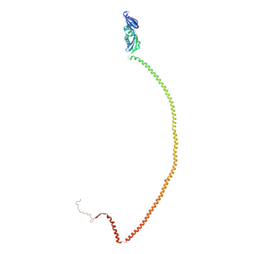
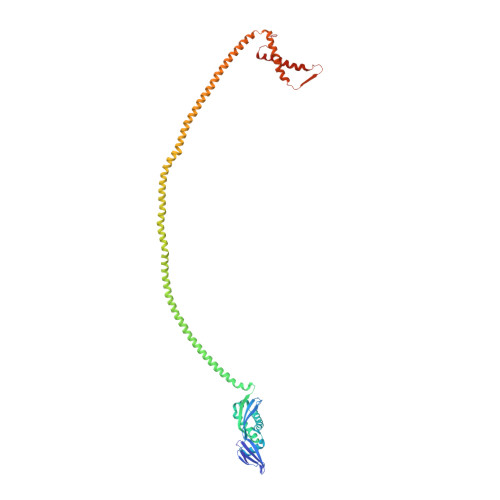
    In this study, we used cryoelectron microscopy to determine the structures of the Flotillin protein complex, part of the Stomatin, Prohibitin, Flotillin, and HflK/C (SPFH) superfamily, from cell-derived vesicles without detergents. It forms a right-handed helical barrel consisting of 22 pairs of Flotillin1 and Flotillin2 subunits, with a diameter of 32 nm at its wider end and 19 nm at its narrower end. Oligomerization is stabilized by the C terminus, which forms two helical layers linked by a β-strand, and coiled-coil domains that enable strong charge-charge intersubunit interactions. Flotillin interacts with membranes at both ends; through its SPFH1 domains at the wide end and the C terminus at the narrow end, facilitated by hydrophobic interactions and lipidation. The inward tilting of the SPFH domain, likely triggered by phosphorylation, suggests its role in membrane curvature induction, which could be connected to its proposed role in clathrin-independent endocytosis. The structure suggests a shared architecture across the family of SPFH proteins and will promote further research into Flotillin's roles in cell biology.


  • Organizational Affiliation

    Laboratory of Molecular Neurobiology and Biophysics, The Rockefeller University, New York, NY 10065.


Macromolecules
Find similar proteins by:  (by identity cutoff)  |  3D Structure
Entity ID: 1
MoleculeChains Sequence LengthOrganismDetailsImage
Flotillin-2402Homo sapiensMutation(s): 0 
UniProt & NIH Common Fund Data Resources
Find proteins for J3QLD9 (Homo sapiens)
Explore J3QLD9 
Go to UniProtKB:  J3QLD9
GTEx:  ENSG00000132589 
Entity Groups  
Sequence Clusters30% Identity50% Identity70% Identity90% Identity95% Identity100% Identity
UniProt GroupJ3QLD9
Sequence Annotations
Expand
  • Reference Sequence
Find similar proteins by:  (by identity cutoff)  |  3D Structure
Entity ID: 2
MoleculeChains Sequence LengthOrganismDetailsImage
Flotillin-1422Homo sapiensMutation(s): 0 
UniProt & NIH Common Fund Data Resources
Find proteins for O75955 (Homo sapiens)
Explore O75955 
Go to UniProtKB:  O75955
PHAROS:  O75955
GTEx:  ENSG00000137312 
Entity Groups  
Sequence Clusters30% Identity50% Identity70% Identity90% Identity95% Identity100% Identity
UniProt GroupO75955
Sequence Annotations
Expand
  • Reference Sequence
Experimental Data & Validation

Experimental Data

  • Method: ELECTRON MICROSCOPY
  • Resolution: 3.50 Å
  • Aggregation State: PARTICLE 
  • Reconstruction Method: SINGLE PARTICLE 

Structure Validation

View Full Validation Report


Entry History & Funding Information

Deposition Data


Funding OrganizationLocationGrant Number
National Institutes of Health/National Institute of General Medical Sciences (NIH/NIGMS)United StatesGM43949
Howard Hughes Medical Institute (HHMI)United States--

Revision History  (Full details and data files)

  • Version 1.0: 2024-07-24
    Type: Initial release