8XSB | pdb_00008xsb

Crystal structure of the DNA-bound AHR-ARNT heterodimer in complex with Indirubin


Experimental Data Snapshot

  • Method: X-RAY DIFFRACTION
  • Resolution: 3.06 Å
  • R-Value Free: 
    0.240 (Depositor), 0.239 (DCC) 
  • R-Value Work: 
    0.200 (Depositor), 0.203 (DCC) 
  • R-Value Observed: 
    0.202 (Depositor) 

Starting Model: experimental
View more details

wwPDB Validation   3D Report Full Report


Ligand Structure Quality Assessment 

Created with Raphaël 2.3.0Worse 01 BetterLigand structure goodness of fit to experimental dataBest fitted JY6Click on this verticalbar to view details

This is version 1.1 of the entry. See complete history


Literature

Structural basis for the ligand-dependent activation of heterodimeric AHR-ARNT complex.

Diao, X.Shang, Q.Guo, M.Huang, Y.Zhang, M.Chen, X.Liang, Y.Sun, X.Zhou, F.Zhuang, J.Liu, S.J.Vogel, C.F.A.Rastinejad, F.Wu, D.

(2025) Nat Commun 16: 1282-1282

  • DOI: https://doi.org/10.1038/s41467-025-56574-7
  • Primary Citation of Related Structures:  
    8XS6, 8XS7, 8XS8, 8XS9, 8XSA, 8XSB

  • PubMed Abstract: 

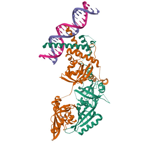
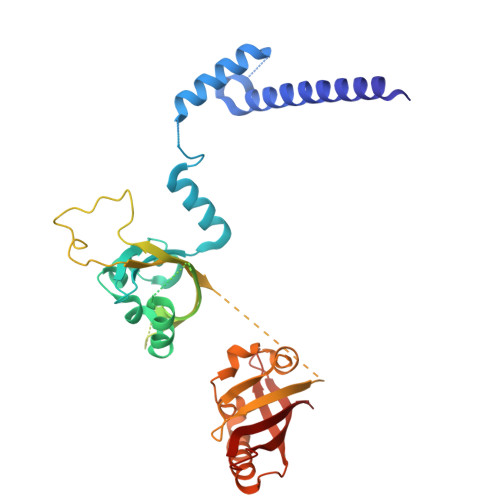
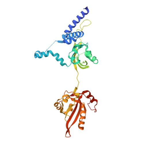
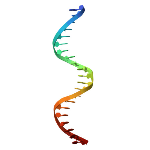
    The aryl hydrocarbon receptor (AHR) possesses an extraordinary capacity to sense and respond to a wide range of small-molecule ligands, ranging from polycyclic aromatic hydrocarbons to endogenous compounds. Upon ligand binding, AHR translocates from the cytoplasm to nucleus, forming a transcriptionally active complex with aryl hydrocarbon receptor nuclear translocator (ARNT), for DNA binding and initiation of gene expression programs that include cellular detoxification pathways and immune responses. Here, we examine the molecular mechanisms governing AHR's high-affinity binding and activation by a diverse group of ligands. Crystal structures of the AHR-ARNT-DNA complexes, bound with each of six established AHR ligands, including Tapinarof, 6-formylindolo[3,2-b]carbazole (FICZ), benzo[a]pyrene (BaP), β-naphthoflavone (BNF), Indigo and Indirubin, reveal an unconventional mode of subunit assembly with intimate association between the PAS-B domains of AHR and ARNT. AHR's PAS-B domain utilizes eight conserved residues whose dynamic rearrangements account for the ability to bind to ligands through hydrophobic and π-π interactions. Our findings further reveal the structural underpinnings of a ligand-driven activation mechanism, whereby a segment of the AHR protein undergoes a structural transition from chaperone engagement to ARNT heterodimer stabilization, to generate the transcriptionally competent assembly. Our results provide key information for the future development of AHR-targeting drugs.


  • Organizational Affiliation

    State Key Laboratory of Microbial Technology, Shandong University, Qingdao, China.


Macromolecules

Find similar proteins by:  (by identity cutoff)  |  3D Structure
Entity ID: 1
MoleculeChains Sequence LengthOrganismDetailsImage
Aryl hydrocarbon receptor nuclear translocator382Homo sapiensMutation(s): 0 
Gene Names: ARNT
UniProt & NIH Common Fund Data Resources
Find proteins for P27540 (Homo sapiens)
Explore P27540 
Go to UniProtKB:  P27540
PHAROS:  P27540
GTEx:  ENSG00000143437 
Entity Groups  
Sequence Clusters30% Identity50% Identity70% Identity90% Identity95% Identity100% Identity
UniProt GroupP27540
Sequence Annotations
Expand
  • Reference Sequence
Find similar proteins by:  (by identity cutoff)  |  3D Structure
Entity ID: 2
MoleculeChains Sequence LengthOrganismDetailsImage
Aryl hydrocarbon receptor395Sus scrofaMutation(s): 0 
Gene Names: AHR
UniProt
Find proteins for I3LF82 (Sus scrofa)
Explore I3LF82 
Go to UniProtKB:  I3LF82
Entity Groups  
Sequence Clusters30% Identity50% Identity70% Identity90% Identity95% Identity100% Identity
UniProt GroupI3LF82
Sequence Annotations
Expand
  • Reference Sequence
Small Molecules
Ligands 1 Unique
IDChains Name / Formula / InChI Key2D Diagram3D Interactions
JY6 (Subject of Investigation/LOI)
Query on JY6

Download Ideal Coordinates CCD File 
E [auth B](3~{Z})-3-(3-oxidanylidene-1~{H}-indol-2-ylidene)-1~{H}-indol-2-one
C16 H10 N2 O2
CRDNMYFJWFXOCH-YPKPFQOOSA-N
Experimental Data & Validation

Experimental Data

  • Method: X-RAY DIFFRACTION
  • Resolution: 3.06 Å
  • R-Value Free:  0.240 (Depositor), 0.239 (DCC) 
  • R-Value Work:  0.200 (Depositor), 0.203 (DCC) 
  • R-Value Observed: 0.202 (Depositor) 
Space Group: P 1 21 1
Unit Cell:
Length ( Å )Angle ( ˚ )
a = 68.1α = 90
b = 99.632β = 90.51
c = 80.002γ = 90
Software Package:
Software NamePurpose
PHENIXrefinement
XDSdata scaling
XDSdata reduction
PHENIXphasing

Structure Validation

View Full Validation Report


Ligand Structure Quality Assessment 

Created with Raphaël 2.3.0Worse 01 BetterLigand structure goodness of fit to experimental dataBest fitted JY6Click on this verticalbar to view details

Entry History & Funding Information

Deposition Data


Funding OrganizationLocationGrant Number
National Natural Science Foundation of China (NSFC)China82373876

Revision History  (Full details and data files)

  • Version 1.0: 2025-01-15
    Type: Initial release
  • Version 1.1: 2025-02-19
    Changes: Database references