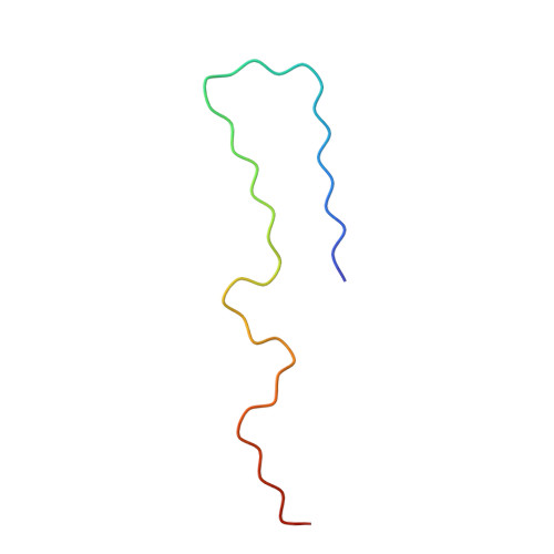
Cryo-EM structures of A beta 40 filaments from the leptomeninges of individuals with Alzheimer's disease and cerebral amyloid angiopathy.
Yang, Y., Murzin, A.G., Peak-Chew, S., Franco, C., Garringer, H.J., Newell, K.L., Ghetti, B., Goedert, M., Scheres, S.H.W.(2023) Acta Neuropathol Commun 11: 191-191
- PubMed: 38049918
- DOI: https://doi.org/10.1186/s40478-023-01694-8
- Primary Citation of Related Structures:
8QN6, 8QN7 - PubMed Abstract:
We used electron cryo-microscopy (cryo-EM) to determine the structures of Aβ40 filaments from the leptomeninges of individuals with Alzheimer's disease and cerebral amyloid angiopathy. In agreement with previously reported structures, which were solved to a resolution of 4.4 Å, we found three types of filaments. However, our new structures, solved to a resolution of 2.4 Å, revealed differences in the sequence assignment that redefine the fold of Aβ40 peptides and their interactions. Filaments are made of pairs of protofilaments, the ordered core of which comprises D1-G38. The different filament types comprise one, two or three protofilament pairs. In each pair, residues H14-G37 of both protofilaments adopt an extended conformation and pack against each other in an anti-parallel fashion, held together by hydrophobic interactions and hydrogen bonds between main chains and side chains. Residues D1-H13 fold back on the adjacent parts of their own chains through both polar and non-polar interactions. There are also several additional densities of unknown identity. Sarkosyl extraction and aqueous extraction gave the same structures. By cryo-EM, parenchymal deposits of Aβ42 and blood vessel deposits of Aβ40 have distinct structures, supporting the view that Alzheimer's disease and cerebral amyloid angiopathy are different Aβ proteinopathies.
Organizational Affiliation:
Medical Research Council Laboratory of Molecular Biology, Cambridge, UK. yyang@mrc-lmb.cam.ac.uk.


