8I2N

The RIPK1 kinase domain in complex with QY7-2B compound


Experimental Data Snapshot

  • Method: X-RAY DIFFRACTION
  • Resolution: 2.29 Å
  • R-Value Free: 0.245 
  • R-Value Work: 0.215 
  • R-Value Observed: 0.217 

Starting Model: experimental
View more details

wwPDB Validation   3D Report Full Report


Ligand Structure Quality Assessment 


This is version 1.1 of the entry. See complete history


Literature

Structure-based development of potent and selective type-II kinase inhibitors of RIPK1.

Qin, Y.Li, D.Qi, C.Xiang, H.Meng, H.Liu, J.Zhou, S.Gong, X.Li, Y.Xu, G.Zu, R.Xie, H.Xu, Y.Xu, G.Zhang, Z.Chen, S.Pan, L.Li, Y.Tan, L.

(2024) Acta Pharm Sin B 14: 319-334

  • DOI: https://doi.org/10.1016/j.apsb.2023.10.021
  • Primary Citation of Related Structures:  
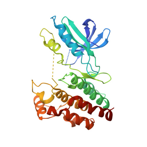
    8I2N

  • PubMed Abstract: 

    Receptor-interacting serine/threonine-protein kinase 1 (RIPK1) functions as a key regulator in inflammation and cell death and is involved in mediating a variety of inflammatory or degenerative diseases. A number of allosteric RIPK1 inhibitors (RIPK1i) have been developed, and some of them have already advanced into clinical evaluation. Recently, selective RIPK1i that interact with both the allosteric pocket and the ATP-binding site of RIPK1 have started to emerge. Here, we report the rational development of a new series of type-II RIPK1i based on the rediscovery of a reported but mechanistically atypical RIPK3i. We also describe the structure-guided lead optimization of a potent, selective, and orally bioavailable RIPK1i, 62 , which exhibits extraordinary efficacies in mouse models of acute or chronic inflammatory diseases. Collectively, 62 provides a useful tool for evaluating RIPK1 in animal disease models and a promising lead for further drug development.


  • Organizational Affiliation

    Interdisciplinary Research Center on Biology and Chemistry, Shanghai Institute of Organic Chemistry, Chinese Academy of Sciences, Shanghai 201210, China.


Macromolecules
Find similar proteins by:  (by identity cutoff)  |  3D Structure
Entity ID: 1
MoleculeChains Sequence LengthOrganismDetailsImage
Receptor-interacting serine/threonine-protein kinase 1
A, B
294Homo sapiensMutation(s): 4 
Gene Names: RIPK1RIPRIP1
EC: 2.7.11.1
UniProt & NIH Common Fund Data Resources
Find proteins for Q13546 (Homo sapiens)
Explore Q13546 
Go to UniProtKB:  Q13546
PHAROS:  Q13546
GTEx:  ENSG00000137275 
Entity Groups  
Sequence Clusters30% Identity50% Identity70% Identity90% Identity95% Identity100% Identity
UniProt GroupQ13546
Sequence Annotations
Expand
  • Reference Sequence
Small Molecules
Ligands 1 Unique
IDChains Name / Formula / InChI Key2D Diagram3D Interactions
O4U (Subject of Investigation/LOI)
Query on O4U

Download Ideal Coordinates CCD File 
C [auth A],
D [auth B]
~{N}-methyl-1-[4-[[[1-methyl-5-(phenylmethyl)pyrazol-3-yl]carbonylamino]methyl]phenyl]benzimidazole-5-carboxamide
C28 H26 N6 O2
OVVBAKUOCIBISQ-UHFFFAOYSA-N
Experimental Data & Validation

Experimental Data

  • Method: X-RAY DIFFRACTION
  • Resolution: 2.29 Å
  • R-Value Free: 0.245 
  • R-Value Work: 0.215 
  • R-Value Observed: 0.217 
  • Space Group: P 21 21 21
Unit Cell:
Length ( Å )Angle ( ˚ )
a = 46.51α = 90
b = 98.742β = 90
c = 128.129γ = 90
Software Package:
Software NamePurpose
PHENIXrefinement
autoPROCdata reduction
autoPROCdata scaling
PHENIXphasing

Structure Validation

View Full Validation Report


Ligand Structure Quality Assessment 


Entry History & Funding Information

Deposition Data


Funding OrganizationLocationGrant Number
National Natural Science Foundation of China (NSFC)China21822705

Revision History  (Full details and data files)

  • Version 1.0: 2024-01-31
    Type: Initial release
  • Version 1.1: 2024-08-21
    Changes: Database references