8CQI

Human heparanase in complex with inhibitor R3794


Experimental Data Snapshot

  • Method: X-RAY DIFFRACTION
  • Resolution: 2.10 Å
  • R-Value Free: 0.253 
  • R-Value Work: 0.199 
  • R-Value Observed: 0.201 

Starting Model: experimental
View more details

wwPDB Validation   3D Report Full Report


Ligand Structure Quality Assessment 


This is version 1.1 of the entry. See complete history


Literature

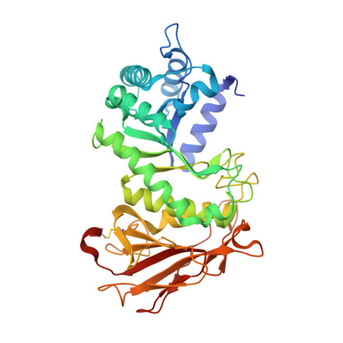
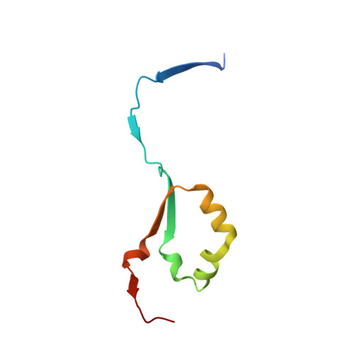
Molecular Basis for Inhibition of Heparanases and beta-Glucuronidases by Siastatin B.

Chen, Y.van den Nieuwendijk, A.M.C.H.Wu, L.Moran, E.Skoulikopoulou, F.van Riet, V.Overkleeft, H.S.Davies, G.J.Armstrong, Z.

(2024) J Am Chem Soc 146: 125-133

  • DOI: https://doi.org/10.1021/jacs.3c04162
  • Primary Citation of Related Structures:  
    8CQI, 8OGX, 8OHQ, 8OHR, 8OHT, 8OHU, 8OHV, 8OHW, 8OHX

  • PubMed Abstract: 

    Siastatin B is a potent and effective iminosugar inhibitor of three diverse glycosidase classes, namely, sialidases, β- d -glucuronidases, and N -acetyl-glucosaminidases. The mode of inhibition of glucuronidases, in contrast to sialidases, has long been enigmatic as siastatin B appears too bulky and incorrectly substituted to be accommodated within a β- d -glucuronidase active site pocket. Herein, we show through crystallographic analysis of protein-inhibitor complexes that siastatin B generates both a hemiaminal and a 3-geminal diol iminosugar (3-GDI) that are, rather than the parent compound, directly responsible for enzyme inhibition. The hemiaminal product is the first observation of a natural product that belongs to the noeuromycin class of inhibitors. Additionally, the 3-GDI represents a new and potent class of the iminosugar glycosidase inhibitor. To substantiate our findings, we synthesized both the gluco - and galacto -configured 3-GDIs and characterized their binding both structurally and kinetically to exo-β- d -glucuronidases and the anticancer target human heparanase. This revealed submicromolar inhibition of exo-β- d -glucuronidases and an unprecedented binding mode by this new class of inhibitor. Our results reveal the mechanism by which siastatin B acts as a broad-spectrum glycosidase inhibitor, identify a new class of glycosidase inhibitor, and suggest new functionalities that can be incorporated into future generations of glycosidase inhibitors.


  • Organizational Affiliation

    Leiden Institute of Chemistry, Leiden University, Einsteinweg 55, 2300 RA Leiden, The Netherlands.


Macromolecules
Find similar proteins by:  (by identity cutoff)  |  3D Structure
Entity ID: 1
MoleculeChains Sequence LengthOrganismDetailsImage
Heparanase 50 kDa subunit389Homo sapiensMutation(s): 1 
Gene Names: HPSEHEPHPAHPA1HPR1HPSE1HSE1
UniProt & NIH Common Fund Data Resources
Find proteins for Q9Y251 (Homo sapiens)
Explore Q9Y251 
Go to UniProtKB:  Q9Y251
PHAROS:  Q9Y251
GTEx:  ENSG00000173083 
Entity Groups  
Sequence Clusters30% Identity50% Identity70% Identity90% Identity95% Identity100% Identity
UniProt GroupQ9Y251
Glycosylation
Glycosylation Sites: 4Go to GlyGen: Q9Y251-1
Sequence Annotations
Expand
  • Reference Sequence
Find similar proteins by:  (by identity cutoff)  |  3D Structure
Entity ID: 2
MoleculeChains Sequence LengthOrganismDetailsImage
Heparanase77Homo sapiensMutation(s): 0 
Gene Names: HPSEHEPHPAHPA1HPR1HPSE1HSE1
EC: 3.2.1.166
UniProt & NIH Common Fund Data Resources
Find proteins for Q9Y251 (Homo sapiens)
Explore Q9Y251 
Go to UniProtKB:  Q9Y251
PHAROS:  Q9Y251
GTEx:  ENSG00000173083 
Entity Groups  
Sequence Clusters30% Identity50% Identity70% Identity90% Identity95% Identity100% Identity
UniProt GroupQ9Y251
Sequence Annotations
Expand
  • Reference Sequence
Small Molecules
Ligands 5 Unique
IDChains Name / Formula / InChI Key2D Diagram3D Interactions
NAG
Query on NAG

Download Ideal Coordinates CCD File 
C [auth A],
D [auth A],
E [auth A],
F [auth A],
K [auth A]
2-acetamido-2-deoxy-beta-D-glucopyranose
C8 H15 N O6
OVRNDRQMDRJTHS-FMDGEEDCSA-N
VGO (Subject of Investigation/LOI)
Query on VGO

Download Ideal Coordinates CCD File 
G [auth A](3~{S},4~{S})-4,5,5-tris(oxidanyl)piperidine-3-carboxylic acid
C6 H11 N O5
VJJFPBNJYGLWRR-IMJSIDKUSA-N
LYY
Query on LYY

Download Ideal Coordinates CCD File 
I [auth A]1,5-anhydro-D-arabinitol
C5 H10 O4
QXAMTEJJAZOINB-QWWZWVQMSA-N
EDO
Query on EDO

Download Ideal Coordinates CCD File 
H [auth A],
J [auth A]
1,2-ETHANEDIOL
C2 H6 O2
LYCAIKOWRPUZTN-UHFFFAOYSA-N
CL
Query on CL

Download Ideal Coordinates CCD File 
L [auth A],
M [auth A],
N [auth A]
CHLORIDE ION
Cl
VEXZGXHMUGYJMC-UHFFFAOYSA-M
Experimental Data & Validation

Experimental Data

  • Method: X-RAY DIFFRACTION
  • Resolution: 2.10 Å
  • R-Value Free: 0.253 
  • R-Value Work: 0.199 
  • R-Value Observed: 0.201 
  • Space Group: P 1 21 1
Unit Cell:
Length ( Å )Angle ( ˚ )
a = 46.769α = 90
b = 71.519β = 95.27
c = 79.32γ = 90
Software Package:
Software NamePurpose
REFMACrefinement
DIALSdata reduction
Aimlessdata scaling
MOLREPphasing

Structure Validation

View Full Validation Report


Ligand Structure Quality Assessment 


Entry History & Funding Information

Deposition Data


Funding OrganizationLocationGrant Number
European Research Council (ERC)European Union--

Revision History  (Full details and data files)

  • Version 1.0: 2024-01-10
    Type: Initial release
  • Version 1.1: 2024-01-24
    Changes: Database references