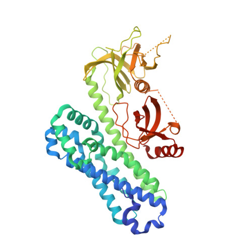
7CSO

Structure of Ephexin4 DH-PH-SH3


Experimental Data Snapshot

  • Method: X-RAY DIFFRACTION
  • Resolution: 2.39 Å
  • R-Value Free: 0.204 
  • R-Value Work: 0.176 
  • R-Value Observed: 0.177 

wwPDB Validation   3D Report Full Report


This is version 1.2 of the entry. See complete history


Literature

Double inhibition and activation mechanisms of Ephexin family RhoGEFs.

Zhang, M.Lin, L.Wang, C.Zhu, J.

(2021) Proc Natl Acad Sci U S A 118

  • DOI: https://doi.org/10.1073/pnas.2024465118
  • Primary Citation of Related Structures:  
    7CSO, 7CSP, 7CSR

  • PubMed Abstract: 

    Ephexin family guanine nucleotide exchange factors (GEFs) transfer signals from Eph tyrosine kinase receptors to Rho GTPases, which play critical roles in diverse cellular processes, as well as cancers and brain disorders. Here, we elucidate the molecular basis underlying inhibition and activation of Ephexin family RhoGEFs. The crystal structures of partially and fully autoinhibited Ephexin4 reveal that the complete autoinhibition requires both N- and C-terminal inhibitory modes, which can operate independently to impede Ras homolog family member G (RhoG) access. This double inhibition mechanism is commonly employed by other Ephexins and SGEF, another RhoGEF for RhoG. Structural, enzymatic, and cell biological analyses show that phosphorylation of a conserved tyrosine residue in its N-terminal inhibitory domain and association of PDZ proteins with its C-terminal PDZ-binding motif may respectively relieve the two autoinhibitory modes in Ephexin4. Our study provides a mechanistic framework for understanding the fine-tuning regulation of Ephexin4 GEF activity and offers possible clues for its pathological dysfunction.


  • Organizational Affiliation

    Ministry of Education Key Laboratory for Membraneless Organelles and Cellular Dynamics, Hefei National Laboratory for Physical Sciences at the Microscale, School of Life Sciences, Division of Life Sciences and Medicine, University of Science and Technology of China, 230027 Hefei, China.


Macromolecules
Find similar proteins by:  (by identity cutoff)  |  3D Structure
Entity ID: 1
MoleculeChains Sequence LengthOrganismDetailsImage
Rho guanine nucleotide exchange factor 16
A, B, C, D
459Mus musculusMutation(s): 0 
Gene Names: Arhgef16
UniProt & NIH Common Fund Data Resources
Find proteins for Q3U5C8 (Mus musculus)
Explore Q3U5C8 
Go to UniProtKB:  Q3U5C8
IMPC:  MGI:2446219
Entity Groups  
Sequence Clusters30% Identity50% Identity70% Identity90% Identity95% Identity100% Identity
UniProt GroupQ3U5C8
Sequence Annotations
Expand
  • Reference Sequence
Small Molecules
Ligands 1 Unique
IDChains Name / Formula / InChI Key2D Diagram3D Interactions
SO4
Query on SO4

Download Ideal Coordinates CCD File 
E [auth A]
F [auth A]
G [auth A]
H [auth B]
I [auth B]
E [auth A],
F [auth A],
G [auth A],
H [auth B],
I [auth B],
J [auth B],
K [auth C],
L [auth C],
M [auth C],
N [auth D],
O [auth D],
P [auth D],
Q [auth D]
SULFATE ION
O4 S
QAOWNCQODCNURD-UHFFFAOYSA-L
Experimental Data & Validation

Experimental Data

  • Method: X-RAY DIFFRACTION
  • Resolution: 2.39 Å
  • R-Value Free: 0.204 
  • R-Value Work: 0.176 
  • R-Value Observed: 0.177 
  • Space Group: P 31 1 2
Unit Cell:
Length ( Å )Angle ( ˚ )
a = 144.444α = 90
b = 144.444β = 90
c = 290.518γ = 120
Software Package:
Software NamePurpose
PHENIXrefinement
XSCALEdata scaling
PDB_EXTRACTdata extraction
XDSdata reduction
CRANK2phasing

Structure Validation

View Full Validation Report


Entry History 

Deposition Data

Revision History  (Full details and data files)

  • Version 1.0: 2021-02-24
    Type: Initial release
  • Version 1.1: 2021-03-03
    Changes: Database references
  • Version 1.2: 2024-03-27
    Changes: Data collection, Database references