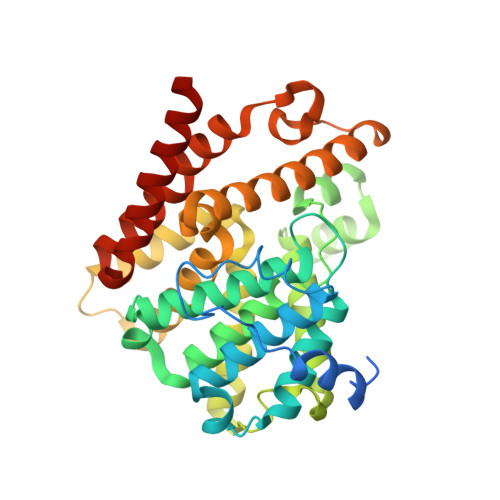
6RFW

Crystal structure of T. brucei PDE-B1 catalytic domain with inhibitor NPD-1039


Experimental Data Snapshot

  • Method: X-RAY DIFFRACTION
  • Resolution: 2.18 Å
  • R-Value Free: 0.234 
  • R-Value Work: 0.175 
  • R-Value Observed: 0.178 

Starting Model: experimental
View more details

wwPDB Validation   3D Report Full Report


Ligand Structure Quality Assessment 


This is version 1.3 of the entry. See complete history


Literature

Alkynamide phthalazinones as a new class of TbrPDEB1 inhibitors (Part 2).

de Heuvel, E.Singh, A.K.Boronat, P.Kooistra, A.J.van der Meer, T.Sadek, P.Blaazer, A.R.Shaner, N.C.Bindels, D.S.Caljon, G.Maes, L.Sterk, G.J.Siderius, M.Oberholzer, M.de Esch, I.J.P.Brown, D.G.Leurs, R.

(2019) Bioorg Med Chem 27: 4013-4029

  • DOI: https://doi.org/10.1016/j.bmc.2019.06.026
  • Primary Citation of Related Structures:  
    6GXQ, 6HWO, 6RB6, 6RCW, 6RFN, 6RFW, 6RGK

  • PubMed Abstract: 

    Inhibitors against Trypanosoma brucei phosphodiesterase B1 (TbrPDEB1) and B2 (TbrPDEB2) have gained interest as new treatments for human African trypanosomiasis. The recently reported alkynamide tetrahydrophthalazinones, which show submicromolar activities against TbrPDEB1 and anti-T. brucei activity, have been used as starting point for the discovery of new TbrPDEB1 inhibitors. Structure-based design indicated that the alkynamide-nitrogen atom can be readily decorated, leading to the discovery of 37, a potent TbrPDEB1 inhibitor with submicromolar activities against T. brucei parasites. Furthermore, 37 is more potent against TbrPDEB1 than hPDE4 and shows no cytotoxicity on human MRC-5 cells. The crystal structures of the catalytic domain of TbrPDEB1 co-crystalized with several different alkynamides show a bidentate interaction with key-residue Gln874, but no interaction with the parasite-specific P-pocket, despite being (uniquely) a more potent inhibitor for the parasite PDE. Incubation of blood stream form trypanosomes by 37 increases intracellular cAMP levels and results in the distortion of the cell cycle and cell death, validating phosphodiesterase inhibition as mode of action.


  • Organizational Affiliation

    Division of Medicinal Chemistry, Amsterdam Institute for Molecules, Medicines and Systems, Vrije Universiteit Amsterdam, 1081 HZ Amsterdam, The Netherlands.


Macromolecules
Find similar proteins by:  (by identity cutoff)  |  3D Structure
Entity ID: 1
MoleculeChains Sequence LengthOrganismDetailsImage
Phosphodiesterase
A, B
360Trypanosoma bruceiMutation(s): 0 
Gene Names: PDEB1
EC: 3.1.4
UniProt
Find proteins for Q8WQX9 (Trypanosoma brucei)
Explore Q8WQX9 
Go to UniProtKB:  Q8WQX9
Entity Groups  
Sequence Clusters30% Identity50% Identity70% Identity90% Identity95% Identity100% Identity
UniProt GroupQ8WQX9
Sequence Annotations
Expand
  • Reference Sequence
Small Molecules
Ligands 6 Unique
IDChains Name / Formula / InChI Key2D Diagram3D Interactions
K1Q (Subject of Investigation/LOI)
Query on K1Q

Download Ideal Coordinates CCD File 
I [auth A],
P [auth B]
3-[5-[(4~{a}~{R},8~{a}~{S})-4-oxidanylidene-3-propan-2-yl-4~{a},5,8,8~{a}-tetrahydrophthalazin-1-yl]-2-methoxy-phenyl]-~{N}-[2-(2-fluorophenyl)ethyl]prop-2-ynamide
C29 H30 F N3 O3
UAIXEGFVIVPDNH-BJKOFHAPSA-N
GOL
Query on GOL

Download Ideal Coordinates CCD File 
G [auth A],
N [auth B],
O [auth B]
GLYCEROL
C3 H8 O3
PEDCQBHIVMGVHV-UHFFFAOYSA-N
ZN
Query on ZN

Download Ideal Coordinates CCD File 
C [auth A],
K [auth B]
ZINC ION
Zn
PTFCDOFLOPIGGS-UHFFFAOYSA-N
GAI
Query on GAI

Download Ideal Coordinates CCD File 
F [auth A],
H [auth A],
J [auth B],
M [auth B]
GUANIDINE
C H5 N3
ZRALSGWEFCBTJO-UHFFFAOYSA-N
FMT
Query on FMT

Download Ideal Coordinates CCD File 
E [auth A]FORMIC ACID
C H2 O2
BDAGIHXWWSANSR-UHFFFAOYSA-N
MG
Query on MG

Download Ideal Coordinates CCD File 
D [auth A],
L [auth B]
MAGNESIUM ION
Mg
JLVVSXFLKOJNIY-UHFFFAOYSA-N
Experimental Data & Validation

Experimental Data

  • Method: X-RAY DIFFRACTION
  • Resolution: 2.18 Å
  • R-Value Free: 0.234 
  • R-Value Work: 0.175 
  • R-Value Observed: 0.178 
  • Space Group: C 1 2 1
Unit Cell:
Length ( Å )Angle ( ˚ )
a = 113.716α = 90
b = 116.553β = 108.17
c = 68.525γ = 90
Software Package:
Software NamePurpose
REFMACrefinement
XDSdata reduction
autoPROCdata scaling
PHASERphasing

Structure Validation

View Full Validation Report


Ligand Structure Quality Assessment 


Entry History & Funding Information

Deposition Data


Funding OrganizationLocationGrant Number
European Union602666

Revision History  (Full details and data files)

  • Version 1.0: 2019-07-24
    Type: Initial release
  • Version 1.1: 2019-08-14
    Changes: Data collection, Database references
  • Version 1.2: 2019-09-04
    Changes: Data collection, Database references
  • Version 1.3: 2024-01-24
    Changes: Data collection, Database references, Derived calculations, Refinement description