6ON9

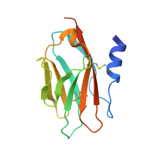
Crystal Structure of the ZIG-8-RIG-5 IG1-IG1 heterodimer, tetragonal form


Experimental Data Snapshot

  • Method: X-RAY DIFFRACTION
  • Resolution: 2.00 Å
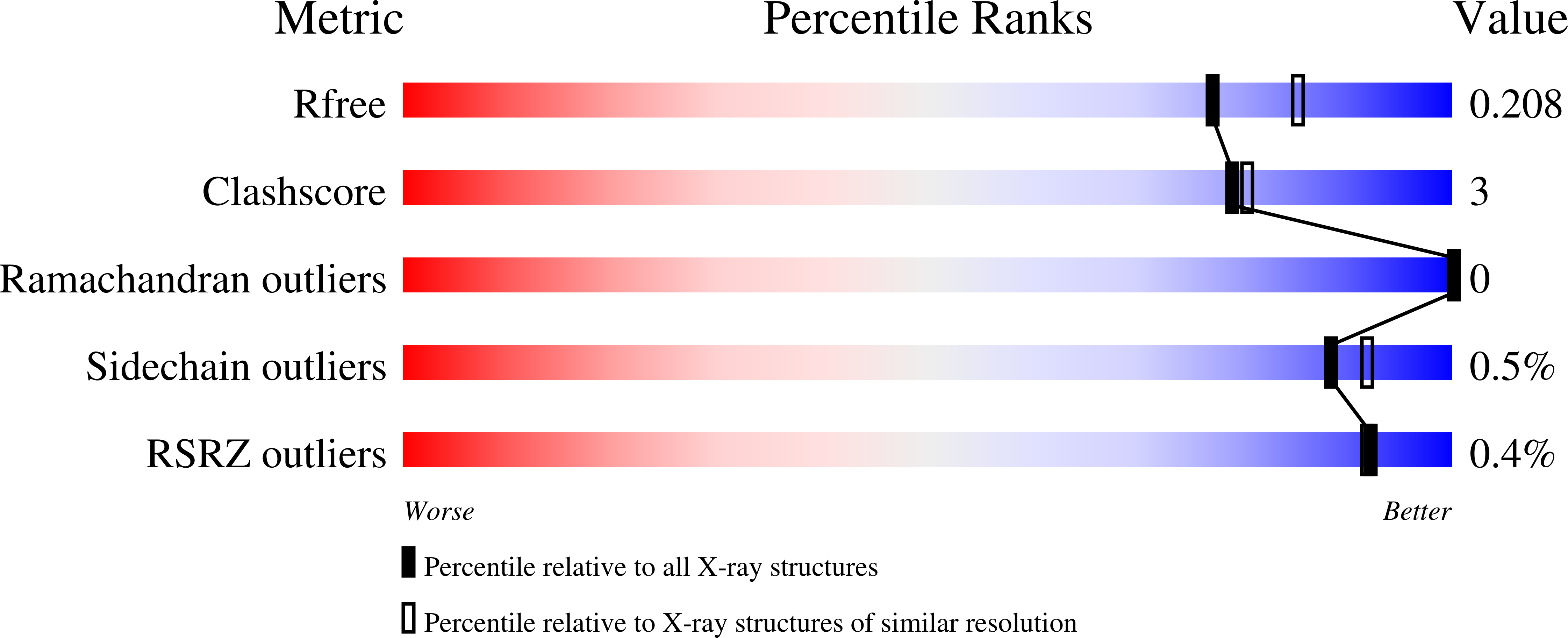
  • R-Value Free: 0.208 
  • R-Value Work: 0.174 
  • R-Value Observed: 0.176 

Starting Model: experimental
View more details

wwPDB Validation   3D Report Full Report


This is version 2.2 of the entry. See complete history


Literature

Family of neural wiring receptors in bilaterians defined by phylogenetic, biochemical, and structural evidence.

Cheng, S.Park, Y.Kurleto, J.D.Jeon, M.Zinn, K.Thornton, J.W.Ozkan, E.

(2019) Proc Natl Acad Sci U S A 116: 9837-9842

  • DOI: https://doi.org/10.1073/pnas.1818631116
  • Primary Citation of Related Structures:  
    6ON6, 6ON9, 6ONB, 6PLL

  • PubMed Abstract: 

    The evolution of complex nervous systems was accompanied by the expansion of numerous protein families, including cell-adhesion molecules, surface receptors, and their ligands. These proteins mediate axonal guidance, synapse targeting, and other neuronal wiring-related functions. Recently, 32 interacting cell surface proteins belonging to two newly defined families of the Ig superfamily (IgSF) in fruit flies were discovered to label different subsets of neurons in the brain and ventral nerve cord. They have been shown to be involved in synaptic targeting and morphogenesis, retrograde signaling, and neuronal survival. Here, we show that these proteins, Dprs and DIPs, are members of a widely distributed family of two- and three-Ig domain molecules with neuronal wiring functions, which we refer to as Wirins. Beginning from a single ancestral Wirin gene in the last common ancestor of Bilateria, numerous gene duplications produced the heterophilic Dprs and DIPs in protostomes, along with two other subfamilies that diversified independently across protostome phyla. In deuterostomes, the ancestral Wirin evolved into the IgLON subfamily of neuronal receptors. We show that IgLONs interact with each other and that their complexes can be broken by mutations designed using homology models based on Dpr and DIP structures. The nematode orthologs ZIG-8 and RIG-5 also form heterophilic and homophilic complexes, and crystal structures reveal numerous apparently ancestral features shared with Dpr-DIP complexes. The evolutionary, biochemical, and structural relationships we demonstrate here provide insights into neural development and the rise of the metazoan nervous system.


  • Organizational Affiliation

    Department of Biochemistry and Molecular Biology, The University of Chicago, Chicago, IL 60637.


Macromolecules
Find similar proteins by:  (by identity cutoff)  |  3D Structure
Entity ID: 1
MoleculeChains Sequence LengthOrganismDetailsImage
Zwei Ig domain protein zig-8125Caenorhabditis elegansMutation(s): 0 
Gene Names: zig-8Y39E4B.8
UniProt
Find proteins for G5ED00 (Caenorhabditis elegans)
Explore G5ED00 
Go to UniProtKB:  G5ED00
Entity Groups  
Sequence Clusters30% Identity50% Identity70% Identity90% Identity95% Identity100% Identity
UniProt GroupG5ED00
Sequence Annotations
Expand
  • Reference Sequence
Find similar proteins by:  (by identity cutoff)  |  3D Structure
Entity ID: 2
MoleculeChains Sequence LengthOrganismDetailsImage
NeuRonal IgCAM-5118Caenorhabditis elegansMutation(s): 0 
Gene Names: rig-5C36F7.4CELE_C36F7.4
UniProt
Find proteins for C6KRM7 (Caenorhabditis elegans)
Explore C6KRM7 
Go to UniProtKB:  C6KRM7
Entity Groups  
Sequence Clusters30% Identity50% Identity70% Identity90% Identity95% Identity100% Identity
UniProt GroupC6KRM7
Glycosylation
Glycosylation Sites: 1
Sequence Annotations
Expand
  • Reference Sequence
Oligosaccharides

Help

Entity ID: 3
MoleculeChains Length2D Diagram Glycosylation3D Interactions
alpha-L-fucopyranose-(1-6)-2-acetamido-2-deoxy-beta-D-glucopyranose
C
2N-Glycosylation
Glycosylation Resources
GlyTouCan:  G86851RC
GlyCosmos:  G86851RC
GlyGen:  G86851RC
Experimental Data & Validation

Experimental Data

Unit Cell:
Length ( Å )Angle ( ˚ )
a = 80.078α = 90
b = 80.078β = 90
c = 166.622γ = 90
Software Package:
Software NamePurpose
XDSdata reduction
PHENIXrefinement
PHASERphasing
XDSdata scaling

Structure Validation

View Full Validation Report


Entry History & Funding Information

Deposition Data


Funding OrganizationLocationGrant Number
National Institutes of Health/National Institute of Neurological Disorders and Stroke (NIH/NINDS)United StatesR01NS097161

Revision History  (Full details and data files)

  • Version 1.0: 2019-05-15
    Type: Initial release
  • Version 1.1: 2019-05-29
    Changes: Data collection, Database references
  • Version 1.2: 2019-12-18
    Changes: Author supporting evidence, Data collection
  • Version 2.0: 2020-07-29
    Type: Remediation
    Reason: Carbohydrate remediation
    Changes: Atomic model, Data collection, Derived calculations, Structure summary
  • Version 2.1: 2023-10-11
    Changes: Data collection, Database references, Derived calculations, Refinement description, Structure summary
  • Version 2.2: 2024-10-30
    Changes: Structure summary