4PRL

Crystal structure of D-lactate dehydrogenase with NAD+ from Lactobacillus jensenii


Experimental Data Snapshot

  • Method: X-RAY DIFFRACTION
  • Resolution: 2.60 Å
  • R-Value Free: 0.278 
  • R-Value Work: 0.211 
  • R-Value Observed: 0.228 

Starting Model: experimental
View more details

wwPDB Validation   3D Report Full Report


Ligand Structure Quality Assessment 


This is version 1.1 of the entry. See complete history


Literature

Crystal structure and thermodynamic properties of d-lactate dehydrogenase from Lactobacillus jensenii.

Kim, S.Gu, S.A.Kim, Y.H.Kim, K.J.

(2014) Int J Biol Macromol 68C: 151-157

  • DOI: https://doi.org/10.1016/j.ijbiomac.2014.04.048
  • Primary Citation of Related Structures:  
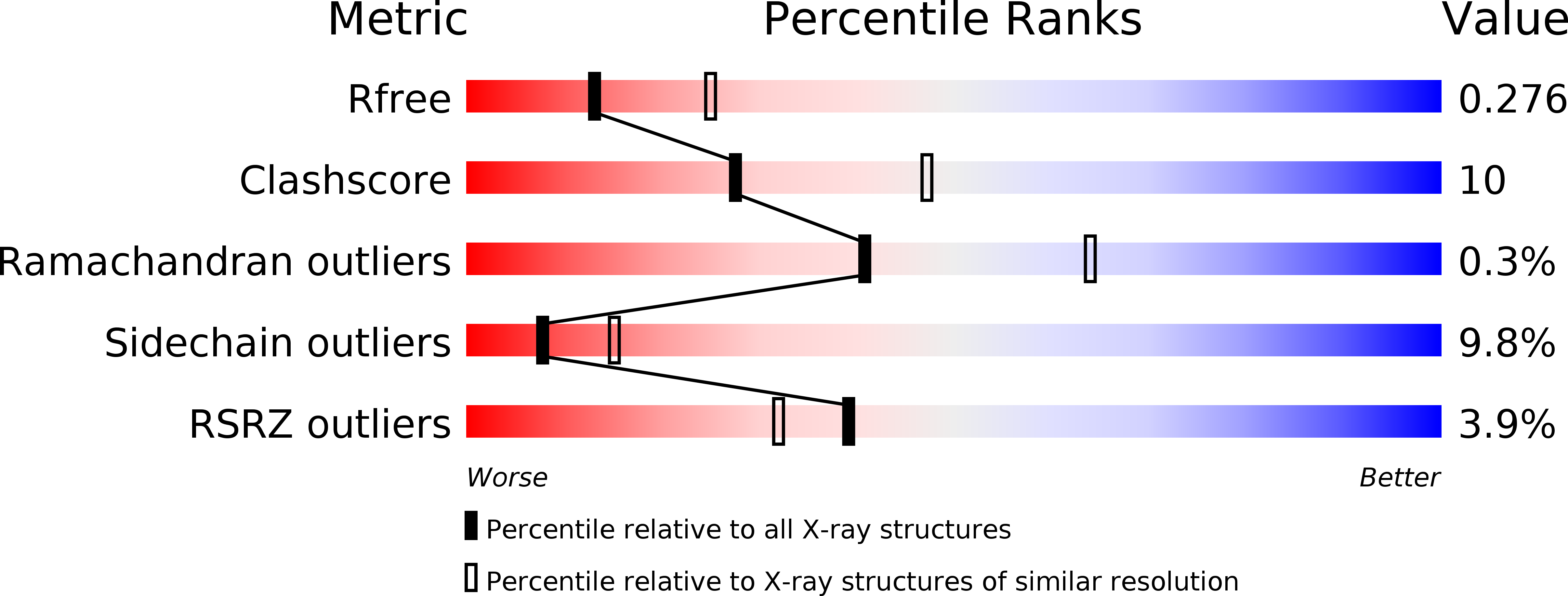
    4PRK, 4PRL

  • PubMed Abstract: 

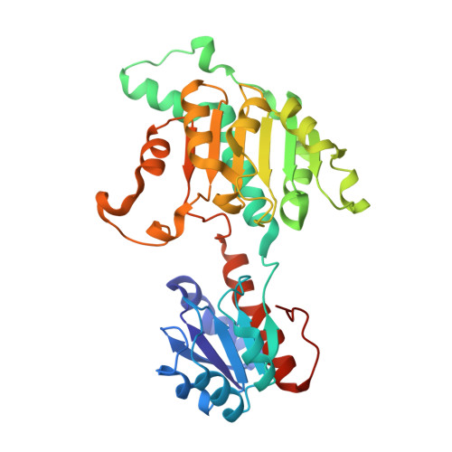
    The thermostable d-lactate dehydrogenase from Lactobacillus jensenii (Ljd-LDH) is a key enzyme in the production of the d-form of lactic acid from pyruvate concomitant with the oxidation of NADH to NAD(+). The polymers of d-lactic acid are used as biodegradable bioplastics. The crystal structures of Ljd-LDH and in complex with NAD(+) were determined at 2.13 and 2.60Å resolutions, respectively. The Ljd-LDH monomer consists of the N-terminal substrate-binding domain and the C-terminal NAD-binding domain. The Ljd-LDH forms a homodimeric structure, and the C-terminal NAD-binding domain mostly enables the dimerization of the enzyme. The NAD cofactor is bound to the GxGxxG NAD-binding motif located between the two domains. Structural comparisons of Ljd-LDH with other d-LDHs reveal that Ljd-LDH has unique amino acid residues at the linker region, which indicates that the open-close dynamics of Ljd-LDH might be different from that of other d-LDHs. Moreover, thermostability experiments showed that the T50(10) value of Ljd-LDH (54.5°C) was much higher than the commercially available d-lactate dehydrogenase (42.7°C). In addition, Ljd-LDH has at least a 7°C higher denaturation temperature compared to commercially available d-LDHs.


  • Organizational Affiliation

    School of Life Sciences, KNU Creative BioResearch Group (BK21 plus program), Kyungpook National University, Daehak-ro 80, Buk-ku, Daegu 702-701, Republic of Korea; School of Nano-Bioscience and Chemical Engineering, Ulsan National Institute of Science and Technology (UNIST), Ulsan 689-798, Republic of Korea.


Macromolecules
Find similar proteins by:  (by identity cutoff)  |  3D Structure
Entity ID: 1
MoleculeChains Sequence LengthOrganismDetailsImage
4-phosphoerythronate dehydrogenase
A, B
336Lactobacillus jensenii 1153Mutation(s): 0 
Gene Names: pdxBLBJG_01197
EC: 1.1.1.290
Entity Groups  
Sequence Clusters30% Identity50% Identity70% Identity90% Identity95% Identity100% Identity
Sequence Annotations
Expand
  • Reference Sequence
Small Molecules
Ligands 1 Unique
IDChains Name / Formula / InChI Key2D Diagram3D Interactions
NAD
Query on NAD

Download Ideal Coordinates CCD File 
C [auth A],
D [auth B]
NICOTINAMIDE-ADENINE-DINUCLEOTIDE
C21 H27 N7 O14 P2
BAWFJGJZGIEFAR-NNYOXOHSSA-N
Experimental Data & Validation

Experimental Data

  • Method: X-RAY DIFFRACTION
  • Resolution: 2.60 Å
  • R-Value Free: 0.278 
  • R-Value Work: 0.211 
  • R-Value Observed: 0.228 
  • Space Group: P 31 2 1
Unit Cell:
Length ( Å )Angle ( ˚ )
a = 90.714α = 90
b = 90.714β = 90
c = 158.183γ = 120
Software Package:
Software NamePurpose
MOLREPphasing
REFMACrefinement
PDB_EXTRACTdata extraction
HKL-2000data collection
HKL-2000data reduction
HKL-2000data scaling

Structure Validation

View Full Validation Report


Ligand Structure Quality Assessment 


Entry History 

Deposition Data

Revision History  (Full details and data files)

  • Version 1.0: 2014-06-18
    Type: Initial release
  • Version 1.1: 2023-11-08
    Changes: Data collection, Database references, Derived calculations, Refinement description