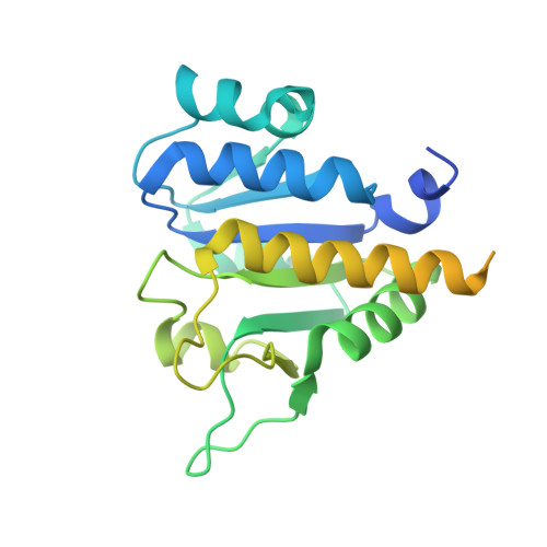
4CND

Crystal structure of E.coli TrmJ


Experimental Data Snapshot

  • Method: X-RAY DIFFRACTION
  • Resolution: 1.50 Å
  • R-Value Free: 0.228 
  • R-Value Work: 0.195 
  • R-Value Observed: 0.197 

Starting Model: experimental
View more details

wwPDB Validation   3D Report Full Report


This is version 1.2 of the entry. See complete history


Literature

Characterization of Two Homologous 2'-O-Methyltransferases Showing Different Specificities for Their tRNA Substrates.

Somme, J.Van Laer, B.Roovers, M.Steyaert, J.Versees, W.Droogmans, L.

(2014) RNA 20: 1257

  • DOI: https://doi.org/10.1261/rna.044503.114
  • Primary Citation of Related Structures:  
    4CND, 4CNE, 4CNF, 4CNG

  • PubMed Abstract: 

    The 2'-O-methylation of the nucleoside at position 32 of tRNA is found in organisms belonging to the three domains of life. Unrelated enzymes catalyzing this modification in Bacteria (TrmJ) and Eukarya (Trm7) have already been identified, but until now, no information is available for the archaeal enzyme. In this work we have identified the methyltransferase of the archaeon Sulfolobus acidocaldarius responsible for the 2'-O-methylation at position 32. This enzyme is a homolog of the bacterial TrmJ. Remarkably, both enzymes have different specificities for the nature of the nucleoside at position 32. While the four canonical nucleosides are substrates of the Escherichia coli enzyme, the archaeal TrmJ can only methylate the ribose of a cytidine. Moreover, the two enzymes recognize their tRNA substrates in a different way. We have solved the crystal structure of the catalytic domain of both enzymes to gain better understanding of these differences at a molecular level.


  • Organizational Affiliation

    Laboratoire de Microbiologie, Université libre de Bruxelles (ULB), 6041 Gosselies, Belgium.


Macromolecules
Find similar proteins by:  (by identity cutoff)  |  3D Structure
Entity ID: 1
MoleculeChains Sequence LengthOrganismDetailsImage
TRNA (CYTIDINE/URIDINE-2'-O-)-METHYLTRANSFERASE TRMJ
A, B
267Escherichia coli K-12Mutation(s): 1 
EC: 2.1.1.200
UniProt
Find proteins for P0AE01 (Escherichia coli (strain K12))
Explore P0AE01 
Go to UniProtKB:  P0AE01
Entity Groups  
Sequence Clusters30% Identity50% Identity70% Identity90% Identity95% Identity100% Identity
UniProt GroupP0AE01
Sequence Annotations
Expand
  • Reference Sequence
Small Molecules
Ligands 1 Unique
IDChains Name / Formula / InChI Key2D Diagram3D Interactions
PEG
Query on PEG

Download Ideal Coordinates CCD File 
C [auth B]DI(HYDROXYETHYL)ETHER
C4 H10 O3
MTHSVFCYNBDYFN-UHFFFAOYSA-N
Experimental Data & Validation

Experimental Data

  • Method: X-RAY DIFFRACTION
  • Resolution: 1.50 Å
  • R-Value Free: 0.228 
  • R-Value Work: 0.195 
  • R-Value Observed: 0.197 
  • Space Group: P 1 21 1
Unit Cell:
Length ( Å )Angle ( ˚ )
a = 42.4α = 90
b = 73.03β = 105.13
c = 53.33γ = 90
Software Package:
Software NamePurpose
REFMACrefinement
XDSdata reduction
XDSdata scaling
PHASERphasing

Structure Validation

View Full Validation Report


Entry History 

Deposition Data

Revision History  (Full details and data files)

  • Version 1.0: 2014-07-02
    Type: Initial release
  • Version 1.1: 2014-07-30
    Changes: Database references
  • Version 1.2: 2023-12-20
    Changes: Data collection, Database references, Derived calculations, Other, Refinement description