4CL2

structure of periplasmic metal binding protein from candidatus liberibacter asiaticus


Experimental Data Snapshot

  • Method: X-RAY DIFFRACTION
  • Resolution: 1.63 Å
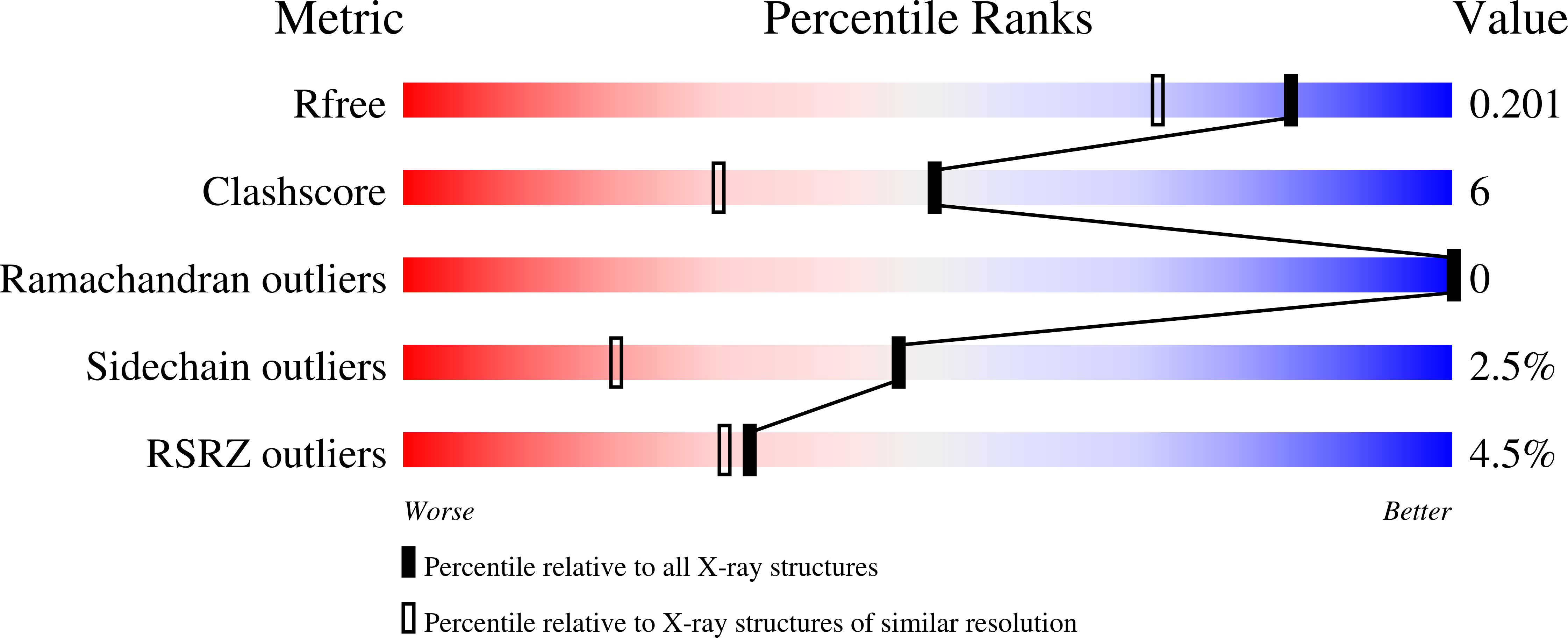
  • R-Value Free: 0.194 
  • R-Value Work: 0.167 
  • R-Value Observed: 0.168 

Starting Model: experimental
View more details

wwPDB Validation   3D Report Full Report


This is version 1.4 of the entry. See complete history


Literature

Crystal Structure of a Periplasmic Solute Binding Protein in Metal-Free, Intermediate and Metal-Bound States from Candidatus Liberibacter Asiaticus.

Sharma, N.Selvakumar, P.Bhose, S.Ghosh, D.K.Kumar, P.Sharma, A.K.

(2015) J Struct Biol 189: 184

  • DOI: https://doi.org/10.1016/j.jsb.2015.01.012
  • Primary Citation of Related Structures:  
    4CL2, 4UDN, 4UDO

  • PubMed Abstract: 

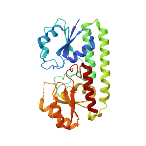
    The Znu system, a member of ABC transporter family, is critical for survival and pathogenesis of Candidatus Liberibacter asiaticus (CLA). Two homologues of this system have been identified in CLA. Here, we report high resolution crystal structure of a periplasmic solute binding protein from second of the two gene clusters of Znu system in CLA (CLas-ZnuA2) in metal-free, intermediate and metal-bound states. CLas-ZnuA2 showed maximum sequence identity to the Mn/Fe-specific solute binding proteins (SBPs) of cluster A-I family. The overall fold of CLas-ZnuA2 is similar to the related cluster A-I family SBPs. The sequence and structure analysis revealed the unique features of CLas-ZnuA2. The comparison of CLas-ZnuA2 structure in three states showed that metal binding and release is facilitated by a large displacement along with a change in orientation of the side chain for one of the metal binding residue (His39) flipped away from metal binding site in metal-free form. The crystal structure captured in intermediate state of metal binding revealed the changes in conformation and interaction of the loop hosting His39 during the metal binding. A rigid body movement of C-domain along with partial unfolding of linker helix at its C-terminal during metal binding, as reported for PsaA, was not observed in CLas-ZnuA2. The present results suggest that despite showing maximum sequence identity to the Mn/Fe-specific SBPs, the mechanistic resemblance of CLas-ZnuA2 seems to be closer to Zn-specific SBPs of cluster A-I family.


  • Organizational Affiliation

    Department of Biotechnology, Indian Institute of Technology Roorkee, Roorkee 247 667, India.


Macromolecules
Find similar proteins by:  (by identity cutoff)  |  3D Structure
Entity ID: 1
MoleculeChains Sequence LengthOrganismDetailsImage
PERIPLASMIC SOLUTE BINDING PROTEIN275Candidatus Liberibacter asiaticus str. psy62Mutation(s): 0 
UniProt
Find proteins for C6XF58 (Liberibacter asiaticus (strain psy62))
Explore C6XF58 
Go to UniProtKB:  C6XF58
Entity Groups  
Sequence Clusters30% Identity50% Identity70% Identity90% Identity95% Identity100% Identity
UniProt GroupC6XF58
Sequence Annotations
Expand
  • Reference Sequence
Small Molecules
Ligands 4 Unique
IDChains Name / Formula / InChI Key2D Diagram3D Interactions
SO4
Query on SO4

Download Ideal Coordinates CCD File 
B [auth A]
C [auth A]
D [auth A]
E [auth A]
F [auth A]
B [auth A],
C [auth A],
D [auth A],
E [auth A],
F [auth A],
G [auth A]
SULFATE ION
O4 S
QAOWNCQODCNURD-UHFFFAOYSA-L
GOL
Query on GOL

Download Ideal Coordinates CCD File 
J [auth A],
K [auth A]
GLYCEROL
C3 H8 O3
PEDCQBHIVMGVHV-UHFFFAOYSA-N
ACT
Query on ACT

Download Ideal Coordinates CCD File 
H [auth A],
I [auth A]
ACETATE ION
C2 H3 O2
QTBSBXVTEAMEQO-UHFFFAOYSA-M
MN
Query on MN

Download Ideal Coordinates CCD File 
L [auth A]MANGANESE (II) ION
Mn
WAEMQWOKJMHJLA-UHFFFAOYSA-N
Experimental Data & Validation

Experimental Data

  • Method: X-RAY DIFFRACTION
  • Resolution: 1.63 Å
  • R-Value Free: 0.194 
  • R-Value Work: 0.167 
  • R-Value Observed: 0.168 
  • Space Group: P 32 2 1
Unit Cell:
Length ( Å )Angle ( ˚ )
a = 94.06α = 90
b = 94.06β = 90
c = 94.51γ = 120
Software Package:
Software NamePurpose
REFMACrefinement
iMOSFLMdata reduction
SCALAdata scaling
MOLREPphasing

Structure Validation

View Full Validation Report


Entry History 

Deposition Data

Revision History  (Full details and data files)

  • Version 1.0: 2015-01-28
    Type: Initial release
  • Version 1.1: 2015-02-11
    Changes: Database references
  • Version 1.2: 2015-03-18
    Changes: Database references
  • Version 1.3: 2018-05-30
    Changes: Data collection
  • Version 1.4: 2023-12-20
    Changes: Data collection, Database references, Derived calculations, Other, Refinement description