3ULR

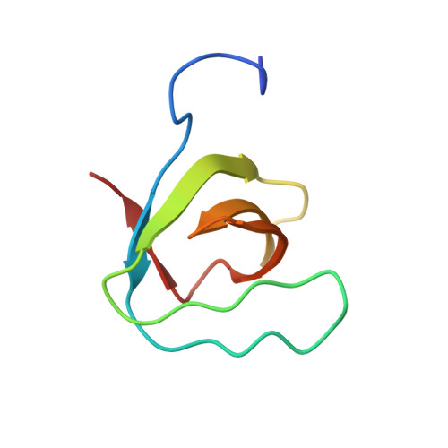
Lysozyme contamination facilitates crystallization of a hetero-trimericCortactin:Arg:Lysozyme complex


Experimental Data Snapshot

  • Method: X-RAY DIFFRACTION
  • Resolution: 1.65 Å
  • R-Value Free: 0.225 
  • R-Value Work: 0.184 
  • R-Value Observed: 0.186 

Starting Models: experimental
View more details

wwPDB Validation   3D Report Full Report


This is version 1.2 of the entry. See complete history


Literature

Lysozyme contamination facilitates crystallization of a heterotrimeric cortactin-Arg-lysozyme complex.

Liu, W.Macgrath, S.M.Koleske, A.J.Boggon, T.J.

(2012) Acta Crystallogr Sect F Struct Biol Cryst Commun 68: 154-158

  • DOI: https://doi.org/10.1107/S1744309111056132
  • Primary Citation of Related Structures:  
    3ULR

  • PubMed Abstract: 

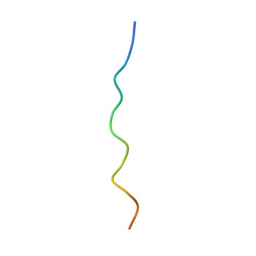
    Crystallization of contaminating proteins is a frequently encountered problem for macromolecular crystallographers. In this study, an attempt was made to obtain a binary cocrystal structure of the SH3 domain of cortactin and a 17-residue peptide from the Arg nonreceptor tyrosine kinase encompassing a PxxPxxPxxP (PxxP1) motif. However, cocrystals could only be obtained in the presence of trace amounts of a contaminating protein. A structure solution obtained by molecular replacement followed by ARP/wARP automatic model building allowed a 'sequence-by-crystallography' approach to discover that the contaminating protein was lysozyme. This 1.65 Å resolution crystal structure determination of a 1:1:1 heterotrimeric complex of Arg, cortactin and lysozyme thus provides an unusual `caveat emptor' warning of the dangers that underpurified proteins harbor for macromolecular crystallographers.


  • Organizational Affiliation

    Department of Pharmacology, Yale University School of Medicine, New Haven, CT 06520, USA.


Macromolecules
Find similar proteins by:  (by identity cutoff)  |  3D Structure
Entity ID: 1
MoleculeChains Sequence LengthOrganismDetailsImage
Lysozyme C129Gallus gallusMutation(s): 0 
Gene Names: LYZ
EC: 3.2.1.17
UniProt
Find proteins for P00698 (Gallus gallus)
Explore P00698 
Go to UniProtKB:  P00698
Entity Groups  
Sequence Clusters30% Identity50% Identity70% Identity90% Identity95% Identity100% Identity
UniProt GroupP00698
Sequence Annotations
Expand
  • Reference Sequence
Find similar proteins by:  (by identity cutoff)  |  3D Structure
Entity ID: 2
MoleculeChains Sequence LengthOrganismDetailsImage
Src substrate cortactin65Mus musculusMutation(s): 0 
Gene Names: CttnEms1
UniProt & NIH Common Fund Data Resources
Find proteins for Q60598 (Mus musculus)
Explore Q60598 
Go to UniProtKB:  Q60598
IMPC:  MGI:99695
Entity Groups  
Sequence Clusters30% Identity50% Identity70% Identity90% Identity95% Identity100% Identity
UniProt GroupQ60598
Sequence Annotations
Expand
  • Reference Sequence

Find similar proteins by:  Sequence   |   3D Structure  

Entity ID: 3
MoleculeChains Sequence LengthOrganismDetailsImage
Abelson tyrosine-protein kinase 217Homo sapiensMutation(s): 0 
EC: 2.7.10.2
UniProt & NIH Common Fund Data Resources
Find proteins for P42684 (Homo sapiens)
Explore P42684 
Go to UniProtKB:  P42684
PHAROS:  P42684
GTEx:  ENSG00000143322 
Entity Groups  
Sequence Clusters30% Identity50% Identity70% Identity90% Identity95% Identity100% Identity
UniProt GroupP42684
Sequence Annotations
Expand
  • Reference Sequence
Experimental Data & Validation

Experimental Data

Unit Cell:
Length ( Å )Angle ( ˚ )
a = 38.658α = 90
b = 57.838β = 90
c = 95.513γ = 90
Software Package:
Software NamePurpose
ADSCdata collection
PHASERphasing
REFMACrefinement
HKL-2000data reduction
HKL-2000data scaling

Structure Validation

View Full Validation Report


Entry History 

Deposition Data

Revision History  (Full details and data files)

  • Version 1.0: 2012-01-11
    Type: Initial release
  • Version 1.1: 2012-02-15
    Changes: Database references
  • Version 1.2: 2023-09-13
    Changes: Data collection, Database references, Refinement description