Catalytic Mechanism of Phosphorylase Kinase Probed by Mutational Studies.
Skamnaki, V.T., Owen, D.J., Noble, M.E., Lowe, E.D., Lowe, G., Oikonomakos, N.G., Johnson, L.N.(1999) Biochemistry 38: 14718
- PubMed: 10545198
- DOI: https://doi.org/10.1021/bi991454f
- Primary Citation of Related Structures:
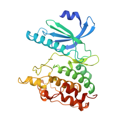
1QL6 - PubMed Abstract:
The contributions to catalysis of the conserved catalytic aspartate (Asp149) in the phosphorylase kinase catalytic subunit (PhK; residues 1-298) have been studied by kinetic and crystallographic methods. Kinetic studies in solvents of different viscosity show that PhK, like cyclic AMP dependent protein kinase, exhibits a mechanism in which the chemical step of phosphoryl transfer is fast and the rate-limiting step is release of the products, ADP and phosphoprotein, and possibly viscosity-dependent conformational changes. Site-directed mutagenesis of Asp149 to Ala and Asn resulted in enzymes with a small increase in K(m) for glycogen phosphorylase b (GPb) and ATP substrates and dramatic decreases in k(cat) (1.3 x 10(4) for Asp149Ala and 4.7 x 10(3) for Asp149Asn mutants, respectively). Viscosometric kinetic measurements with the Asp149Asn mutant showed a reduction in the rate-limiting step for release of products by 4.5 x 10(3) and a significant decrease (possibly as great as 2.2 x 10(3)) in the rate constant characterizing the chemical step. The date combined with the crystallographic evidence for the ternary PhK-AMPPNP-peptide complex [Lowe et al. (1997) EMBO J. 6, 6646-6658] provide powerful support for the role of the carboxyl of Asp149 in binding and orientation of the substrate and in catalysis of phosphoryl transfer. The constitutively active subunit PhK has a glutamate (Glu182) residue in the activation segment, in place of a phosphorylatable serine, threonine, or tyrosine residue in other protein kinases that are activated by phosphorylation. Site-directed mutagenesis of Glu182 and other residues involved in a hydrogen bond network resulted in mutant proteins (Glu182Ser, Arg148Ala, and Tyr206Phe) with decreased catalytic efficiency (approximate average decrease in k(cat)/K(m) by 20-fold). The crystal structure of the mutant Glu182Ser at 2.6 A resolution showed a phosphate dianion about 2.6 A from the position previously occupied by the carboxylate of Glu182. There was no change in tertiary structure from the native protein, but the activation segment in the region C-terminal to residue 182 showed increased disorder, indicating that correct localization of the activation segment is necessary in order to recognize and present the protein substrate for catalysis.
Organizational Affiliation:
Institute for Biological Research and Biotechnology, Athens, Greece.