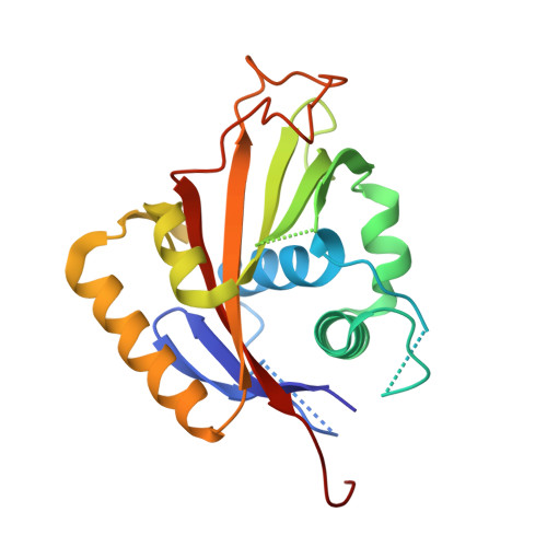
RCSB PDB - 1Q3I: Crystal Structure of Na,K-ATPase N-domain

 1Q3I

Crystal Structure of Na,K-ATPase N-domain


Experimental Data Snapshot

  • Method: X-RAY DIFFRACTION
  • Resolution: 2.60 Å
  • R-Value Free: 0.292 
  • R-Value Work: 0.251 

wwPDB Validation   3D Report Full Report


This is version 1.5 of the entry. See complete history


Literature

The Crystallographic Structure of Na,K-ATPase N-domain at 2.6 A Resolution

Hakansson, K.O.

(2003) J Mol Biol 332: 1175-1182

  • DOI: https://doi.org/10.1016/j.jmb.2003.07.012
  • Primary Citation of Related Structures:  
    1Q3I

  • PubMed Abstract: 

    The structure of the N-domain of porcine alpha(2) Na,K-ATPase was determined crystallographically to 3.2A resolution by isomorphous heavy-atom replacement using a single mercury derivative. The structure was finally refined against 2.6A resolution synchrotron data. The domain forms a seven-stranded antiparallel beta-sheet with two additional beta-strands forming a hairpin and five alpha-helices. Approximately 75% of the residues were superimposable with residues from the structure of Ca-ATPase N-domain, and a structure-based sequence alignment is presented. The positions of key residues are discussed in relation to the pattern of hydrophobicity, charge and sequence conservation of the molecular surface. The structure of a hexahistidine tag binding to nickel ions is presented.


  • Organizational Affiliation

    August Krogh Institute, Copenhagen University, Universitetsparken 13, DK-2100 OE, Copenhagen, Denmark. kohakansson@aki.ku.dk


Macromolecules
Find similar proteins by:  (by identity cutoff)  |  3D Structure
Entity ID: 1
MoleculeChains Sequence LengthOrganismDetailsImage
Na,K-ATPase214Sus scrofaMutation(s): 0 
EC: 3.6.3.9 (PDB Primary Data), 7.2.2.13 (UniProt)
UniProt
Find proteins for D2WKD8 (Sus scrofa)
Explore D2WKD8 
Go to UniProtKB:  D2WKD8
Entity Groups  
Sequence Clusters30% Identity50% Identity70% Identity90% Identity95% Identity100% Identity
UniProt GroupD2WKD8
Sequence Annotations
Expand
  • Reference Sequence
Experimental Data & Validation

Experimental Data

  • Method: X-RAY DIFFRACTION
  • Resolution: 2.60 Å
  • R-Value Free: 0.292 
  • R-Value Work: 0.251 
  • Space Group: F 2 3
Unit Cell:
Length ( Å )Angle ( ˚ )
a = 147.31α = 90
b = 147.31β = 90
c = 147.31γ = 90
Software Package:
Software NamePurpose
MOSFLMdata reduction
SCALAdata scaling
CNSrefinement
CCP4data scaling
CNSphasing

Structure Validation

View Full Validation Report


Entry History 

Deposition Data

Revision History  (Full details and data files)

  • Version 1.0: 2004-08-03
    Type: Initial release
  • Version 1.1: 2008-04-29
    Changes: Version format compliance
  • Version 1.2: 2011-07-13
    Changes: Derived calculations, Version format compliance
  • Version 1.3: 2011-11-16
    Changes: Atomic model
  • Version 1.4: 2018-03-07
    Changes: Data collection
  • Version 1.5: 2024-02-14
    Changes: Data collection, Database references, Derived calculations