Kinetic Resolution of the Atomic 3D Structures Formed by Ground and Excited Conformational States in an RNA Dynamic Ensemble.
Roy, R., Geng, A., Shi, H., Merriman, D.K., Dethoff, E.A., Salmon, L., Al-Hashimi, H.M.(2023) J Am Chem Soc 145: 22964-22978
- PubMed: 37831584
- DOI: https://doi.org/10.1021/jacs.3c04614
- Primary Citation of Related Structures:
8THV - PubMed Abstract:
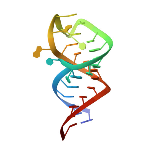
Knowing the 3D structures formed by the various conformations populating the RNA free-energy landscape, their relative abundance, and kinetic interconversion rates is required to obtain a quantitative and predictive understanding of how RNAs fold and function at the atomic level. While methods integrating ensemble-averaged experimental data with computational modeling are helping define the most abundant conformations in RNA ensembles, elucidating their kinetic rates of interconversion and determining the 3D structures of sparsely populated short-lived RNA excited conformational states (ESs) remains challenging. Here, we developed an approach integrating Rosetta-FARFAR RNA structure prediction with NMR residual dipolar couplings and relaxation dispersion that simultaneously determines the 3D structures formed by the ground-state (GS) and ES subensembles, their relative abundance, and kinetic rates of interconversion. The approach is demonstrated on HIV-1 TAR, whose six-nucleotide apical loop was previously shown to form a sparsely populated (∼13%) short-lived (lifetime ∼ 45 μs) ES. In the GS, the apical loop forms a broad distribution of open conformations interconverting on the pico-to-nanosecond time scale. Most residues are unpaired and preorganized to bind the Tat-superelongation protein complex. The apical loop zips up in the ES, forming a narrow distribution of closed conformations, which sequester critical residues required for protein recognition. Our work introduces an approach for determining the 3D ensemble models formed by sparsely populated RNA conformational states, provides a rare atomic view of an RNA ES, and kinetically resolves the atomic 3D structures of RNA conformational substates, interchanging on time scales spanning 6 orders of magnitude, from picoseconds to microseconds.
Organizational Affiliation:
Center for Genomic and Computational Biology, Duke University School of Medicine, Durham, North Carolina 27710, United States.