A structural rationale for reversible vs irreversible amyloid fibril formation from a single protein.
Frey, L., Zhou, J., Cereghetti, G., Weber, M.E., Rhyner, D., Pokharna, A., Wenchel, L., Kadavath, H., Cao, Y., Meier, B.H., Peter, M., Greenwald, J., Riek, R., Mezzenga, R.(2024) Nat Commun 15: 8448-8448
- PubMed: 39349464
- DOI: https://doi.org/10.1038/s41467-024-52681-z
- Primary Citation of Related Structures:
8QUT, 8QV8 - PubMed Abstract:
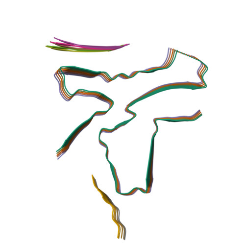
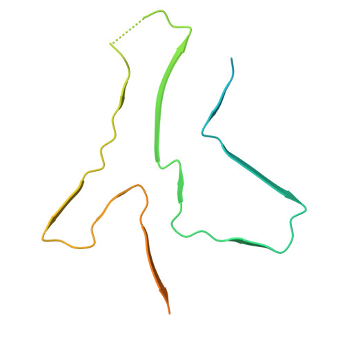
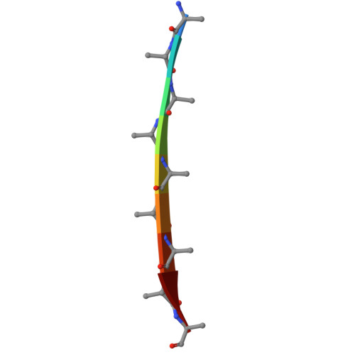
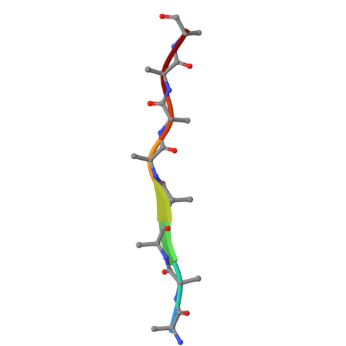
Reversible and irreversible amyloids are two diverging cases of protein (mis)folding associated with the cross-β motif in the protein folding and aggregation energy landscape. Yet, the molecular origins responsible for the formation of reversible vs irreversible amyloids have remained unknown. Here we provide evidence at the atomic level of distinct folding motifs for irreversible and reversible amyloids derived from a single protein sequence: human lysozyme. We compare the 2.8 Å structure of irreversible amyloid fibrils determined by cryo-electron microscopy helical reconstructions with molecular insights gained by solid-state NMR spectroscopy on reversible amyloids. We observe a canonical cross-β-sheet structure in irreversible amyloids, whereas in reversible amyloids, there is a less-ordered coexistence of β-sheet and helical secondary structures that originate from a partially unfolded lysozyme, thus carrying a "memory" of the original folded protein precursor. We also report the structure of hen egg-white lysozyme irreversible amyloids at 3.2 Å resolution, revealing another canonical amyloid fold, and reaffirming that irreversible amyloids undergo a complete conversion of the native protein into the cross-β structure. By combining atomic force microscopy, cryo-electron microscopy and solid-state NMR, we show that a full unfolding of the native protein precursor is a requirement for establishing irreversible amyloid fibrils.
Organizational Affiliation:
Institute of Molecular Physical Science, ETH Zürich, Vladimir-Prelog-Weg 2, Zürich, Switzerland.