7NX3 | pdb_00007nx3

Crystal structure of ALK in complex with Fab324


Experimental Data Snapshot

  • Method: X-RAY DIFFRACTION
  • Resolution: 2.81 Å
  • R-Value Free: 
    0.266 (Depositor), 0.260 (DCC) 
  • R-Value Work: 
    0.228 (Depositor), 0.230 (DCC) 
  • R-Value Observed: 
    0.229 (Depositor) 

Starting Model: experimental
View more details

wwPDB Validation   3D Report Full Report


Ligand Structure Quality Assessment 

Created with Raphaël 2.3.0Worse 01 BetterLigand structure goodness of fit to experimental dataBest fitted NAGClick on this verticalbar to view details

This is version 1.4 of the entry. See complete history


Literature

Structural basis of cytokine-mediated activation of ALK family receptors.

De Munck, S.Provost, M.Kurikawa, M.Omori, I.Mukohyama, J.Felix, J.Bloch, Y.Abdel-Wahab, O.Bazan, J.F.Yoshimi, A.Savvides, S.N.

(2021) Nature 600: 143-147

  • DOI: https://doi.org/10.1038/s41586-021-03959-5
  • Primary Citation of Related Structures:  
    7NWZ, 7NX0, 7NX1, 7NX2, 7NX3, 7NX4

  • PubMed Abstract: 

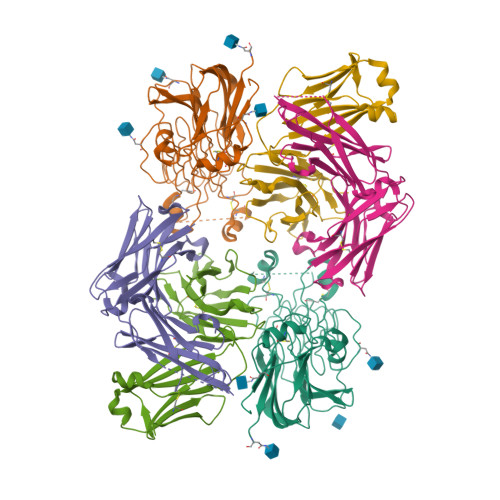
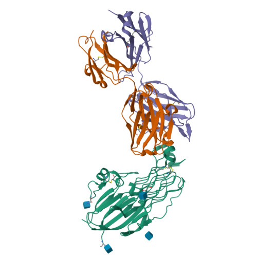
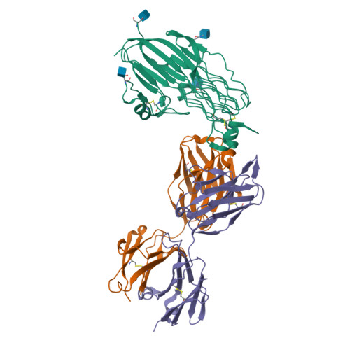
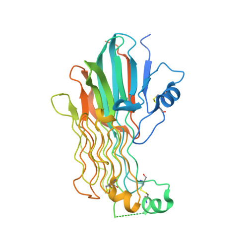
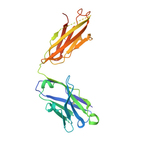
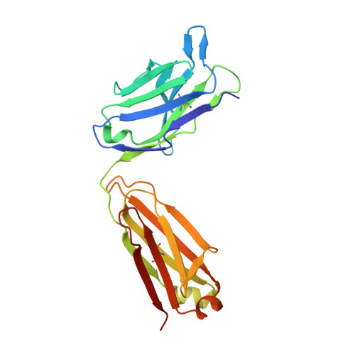
    Anaplastic lymphoma kinase (ALK) 1 and the related leukocyte tyrosine kinase (LTK) 2 are recently deorphanized receptor tyrosine kinases 3 . Together with their activating cytokines, ALKAL1 and ALKAL2 4-6 (also called FAM150A and FAM150B or AUGβ and AUGα, respectively), they are involved in neural development 7 , cancer 7-9 and autoimmune diseases 10 . Furthermore, mammalian ALK recently emerged as a key regulator of energy expenditure and weight gain 11 , consistent with a metabolic role for Drosophila ALK 12 . Despite such functional pleiotropy and growing therapeutic relevance 13,14 , structural insights into ALK and LTK and their complexes with cognate cytokines have remained scarce. Here we show that the cytokine-binding segments of human ALK and LTK comprise a novel architectural chimera of a permuted TNF-like module that braces a glycine-rich subdomain featuring a hexagonal lattice of long polyglycine type II helices. The cognate cytokines ALKAL1 and ALKAL2 are monomeric three-helix bundles, yet their binding to ALK and LTK elicits similar dimeric assemblies with two-fold symmetry, that tent a single cytokine molecule proximal to the cell membrane. We show that the membrane-proximal EGF-like domain dictates the apparent cytokine preference of ALK. Assisted by these diverse structure-function findings, we propose a structural and mechanistic blueprint for complexes of ALK family receptors, and thereby extend the repertoire of ligand-mediated dimerization mechanisms adopted by receptor tyrosine kinases.


  • Organizational Affiliation

    Unit for Structural Biology, Department of Biochemistry and Microbiology, Ghent University, Ghent, Belgium.


Macromolecules
Find similar proteins by:  (by identity cutoff)  |  3D Structure
Entity ID: 1
MoleculeChains Sequence LengthOrganismDetailsImage
ALK tyrosine kinase receptor
A, F
389Homo sapiensMutation(s): 0 
Gene Names: ALK
EC: 2.7.10.1
UniProt & NIH Common Fund Data Resources
Find proteins for Q9UM73 (Homo sapiens)
Explore Q9UM73 
Go to UniProtKB:  Q9UM73
PHAROS:  Q9UM73
GTEx:  ENSG00000171094 
Entity Groups  
Sequence Clusters30% Identity50% Identity70% Identity90% Identity95% Identity100% Identity
UniProt GroupQ9UM73
Glycosylation
Glycosylation Sites: 3Go to GlyGen: Q9UM73-1
Sequence Annotations
Expand
  • Reference Sequence
Find similar proteins by:  (by identity cutoff)  |  3D Structure
Entity ID: 2
MoleculeChains Sequence LengthOrganismDetailsImage
Fab324 HeavyChain
B, E
231Mus musculusMutation(s): 0 
Entity Groups  
Sequence Clusters30% Identity50% Identity70% Identity90% Identity95% Identity100% Identity
Sequence Annotations
Expand
  • Reference Sequence
Find similar proteins by:  (by identity cutoff)  |  3D Structure
Entity ID: 3
MoleculeChains Sequence LengthOrganismDetailsImage
Fab324 Light Chain
C, D
218Mus musculusMutation(s): 0 
Entity Groups  
Sequence Clusters30% Identity50% Identity70% Identity90% Identity95% Identity100% Identity
Sequence Annotations
Expand
  • Reference Sequence
Experimental Data & Validation

Experimental Data

  • Method: X-RAY DIFFRACTION
  • Resolution: 2.81 Å
  • R-Value Free:  0.266 (Depositor), 0.260 (DCC) 
  • R-Value Work:  0.228 (Depositor), 0.230 (DCC) 
  • R-Value Observed: 0.229 (Depositor) 
Space Group: P 21 21 21
Unit Cell:
Length ( Å )Angle ( ˚ )
a = 109.7α = 90
b = 137.47β = 90
c = 144.26γ = 90
Software Package:
Software NamePurpose
PHENIXrefinement
PHENIXrefinement
XDSdata reduction
XDSdata scaling
PHASERphasing

Structure Validation

View Full Validation Report


Ligand Structure Quality Assessment 

Created with Raphaël 2.3.0Worse 01 BetterLigand structure goodness of fit to experimental dataBest fitted NAGClick on this verticalbar to view details

Entry History & Funding Information

Deposition Data


Funding OrganizationLocationGrant Number
Research Foundation - Flanders (FWO)Belgium--

Revision History  (Full details and data files)

  • Version 1.0: 2021-10-27
    Type: Initial release
  • Version 1.1: 2021-11-03
    Changes: Derived calculations
  • Version 1.2: 2021-12-15
    Changes: Database references
  • Version 1.3: 2024-01-31
    Changes: Data collection, Refinement description
  • Version 1.4: 2024-10-09
    Changes: Structure summary