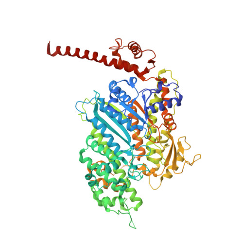
4BYF

Crystal structure of human Myosin 1c in complex with calmodulin in the pre-power stroke state


Experimental Data Snapshot

  • Method: X-RAY DIFFRACTION
  • Resolution: 2.74 Å
  • R-Value Free: 0.237 
  • R-Value Work: 0.183 

Starting Model: experimental
View more details

wwPDB Validation   3D Report Full Report


Ligand Structure Quality Assessment 


This is version 1.2 of the entry. See complete history


Literature

Crystal Structure of Human Myosin 1C-the Motor in Glut4 Exocytosis: Implications for Ca(2+) Regulation and 14-3-3 Binding.

Munnich, S.Taft, M.H.Manstein, D.J.

(2014) J Mol Biol 426: 2070

  • DOI: https://doi.org/10.1016/j.jmb.2014.03.004
  • Primary Citation of Related Structures:  
    4BYF

  • PubMed Abstract: 

    Myosin 1c (Myo1c) plays a key role in supporting motile events that underlie cell migration, vesicle trafficking, insulin-stimulated glucose uptake and hearing. Here, we present the crystal structure of the human Myo1c motor in complex with its light chain calmodulin. Our structure reveals tight interactions of the motor domain with calmodulin bound to the first IQ motif in the neck region. Several of the calmodulin residues contributing to this interaction are also involved in Ca(2+) binding. Contact residues in the motor domain are linked to the central β-sheet and the HO helix, suggesting a mechanism for communicating changes in Ca(2+) binding in the neck region to the actin and nucleotide binding regions of the motor domain. The structural context and the chemical environment of Myo1c mutations that are involved in sensorineural hearing loss in humans are described and their impact on motor function is discussed. We show that a construct consisting of the motor domain of Myo1c and the first IQ motif is sufficient to establish a tight interaction with 14-3-3β (KD=0.9 μM) and present the model of a double-headed Myo1c-14-3-3 complex. This complex has been implicated in the exocytosis of glucose transporter 4 storage vesicles during insulin-stimulated glucose uptake.


  • Organizational Affiliation

    Institute for Biophysical Chemistry, Hannover Medical School, Carl-Neuberg-Str. 1, 30625 Hannover, Germany.


Macromolecules
Find similar proteins by:  (by identity cutoff)  |  3D Structure
Entity ID: 1
MoleculeChains Sequence LengthOrganismDetailsImage
UNCONVENTIONAL MYOSIN-IC
A, C
725Homo sapiensMutation(s): 0 
EC: 3.6.4.1
UniProt & NIH Common Fund Data Resources
Find proteins for O00159 (Homo sapiens)
Explore O00159 
Go to UniProtKB:  O00159
PHAROS:  O00159
GTEx:  ENSG00000197879 
Entity Groups  
Sequence Clusters30% Identity50% Identity70% Identity90% Identity95% Identity100% Identity
UniProt GroupO00159
Sequence Annotations
Expand
  • Reference Sequence
Find similar proteins by:  (by identity cutoff)  |  3D Structure
Entity ID: 2
MoleculeChains Sequence LengthOrganismDetailsImage
CALMODULIN
B, D
149Homo sapiensMutation(s): 0 
UniProt & NIH Common Fund Data Resources
Find proteins for P0DP23 (Homo sapiens)
Explore P0DP23 
Go to UniProtKB:  P0DP23
PHAROS:  P0DP23
GTEx:  ENSG00000198668 
Entity Groups  
Sequence Clusters30% Identity50% Identity70% Identity90% Identity95% Identity100% Identity
UniProt GroupP0DP23
Sequence Annotations
Expand
  • Reference Sequence
Experimental Data & Validation

Experimental Data

  • Method: X-RAY DIFFRACTION
  • Resolution: 2.74 Å
  • R-Value Free: 0.237 
  • R-Value Work: 0.183 
  • Space Group: P 1 21 1
Unit Cell:
Length ( Å )Angle ( ˚ )
a = 59.58α = 90
b = 158.45β = 91.56
c = 114.34γ = 90
Software Package:
Software NamePurpose
PHENIXrefinement
XDSdata reduction
XDSdata scaling
PHASERphasing

Structure Validation

View Full Validation Report


Ligand Structure Quality Assessment 


Entry History 

Deposition Data

Revision History  (Full details and data files)

  • Version 1.0: 2014-03-26
    Type: Initial release
  • Version 1.1: 2014-05-14
    Changes: Database references
  • Version 1.2: 2023-12-20
    Changes: Data collection, Database references, Derived calculations, Other, Refinement description