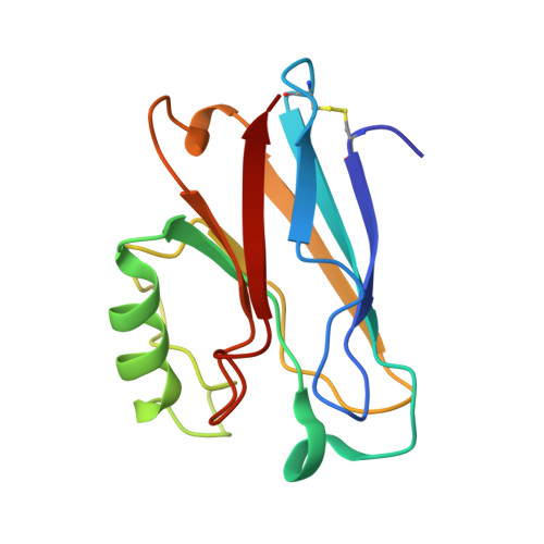
Rationally tuning the reduction potential of a single cupredoxin beyond the natural range.
Marshall, N.M., Garner, D.K., Wilson, T.D., Gao, Y.G., Robinson, H., Nilges, M.J., Lu, Y.(2009) Nature 462: 113-116
- PubMed: 19890331
- DOI: https://doi.org/10.1038/nature08551
- Primary Citation of Related Structures:
3IN0, 3IN2, 3JT2, 3JTB - PubMed Abstract:
Redox processes are at the heart of numerous functions in chemistry and biology, from long-range electron transfer in photosynthesis and respiration to catalysis in industrial and fuel cell research. These functions are accomplished in nature by only a limited number of redox-active agents. A long-standing issue in these fields is how redox potentials are fine-tuned over a broad range with little change to the redox-active site or electron-transfer properties. Resolving this issue will not only advance our fundamental understanding of the roles of long-range, non-covalent interactions in redox processes, but also allow for design of redox-active proteins having tailor-made redox potentials for applications such as artificial photosynthetic centres or fuel cell catalysts for energy conversion. Here we show that two important secondary coordination sphere interactions, hydrophobicity and hydrogen-bonding, are capable of tuning the reduction potential of the cupredoxin azurin over a 700 mV range, surpassing the highest and lowest reduction potentials reported for any mononuclear cupredoxin, without perturbing the metal binding site beyond what is typical for the cupredoxin family of proteins. We also demonstrate that the effects of individual structural features are additive and that redox potential tuning of azurin is now predictable across the full range of cupredoxin potentials.
Organizational Affiliation:
Department of Chemistry, University of Illinois, Urbana-Champaign, Illinois 61801, USA.


