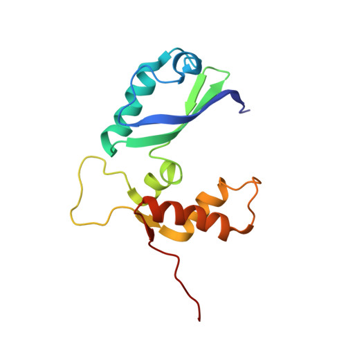
The structure of free L11 and functional dynamics of L11 in free, L11-rRNA(58 nt) binary and L11-rRNA(58 nt)-thiostrepton ternary complexes
Lee, D., Walsh, J.D., Yu, P., Markus, M.A., Choli-Papadopoulou, T., Schwieters, C.D., Krueger, S., Draper, D.E., Wang, Y.X.(2007) J Mol Biol 367: 1007-1022
- PubMed: 17292917
- DOI: https://doi.org/10.1016/j.jmb.2007.01.013
- Primary Citation of Related Structures:
2E34, 2E35, 2E36, 2H8W - PubMed Abstract:
The L11 binding site is one of the most important functional sites in the ribosome. The N-terminal domain of L11 has been implicated as a "reversible switch" in facilitating the coordinated movements associated with EF-G-driven GTP hydrolysis. The reversible switch mechanism has been hypothesized to require conformational flexibility involving re-orientation and re-positioning of the two L11 domains, and warrants a close examination of the structure and dynamics of L11. Here we report the solution structure of free L11, and relaxation studies of free L11, L11 complexed to its 58 nt RNA recognition site, and L11 in a ternary complex with the RNA and thiostrepton antibiotic. The binding site of thiostrepton on L11 was also defined by analysis of structural and dynamics data and chemical shift mapping. The conclusions of this work are as follows: first, the binding of L11 to RNA leads to sizable conformation changes in the regions flanking the linker and in the hinge area that links a beta-sheet and a 3(10)-helix-turn-helix element in the N terminus. Concurrently, the change in the relative orientation may lead to re-positioning of the N terminus, as implied by a decrease of radius of gyration from 18.5 A to 16.2 A. Second, the regions, which undergo large conformation changes, exhibit motions on milliseconds-microseconds or nanoseconds-picoseconds time scales. Third, binding of thiostrepton results in more rigid conformations near the linker (Thr71) and near its putative binding site (Leu12). Lastly, conformational changes in the putative thiostrepton binding site are implicated by the re-emergence of cross-correlation peaks in the spectrum of the ternary complex, which were missing in that of the binary complex. Our combined analysis of both the chemical shift perturbation and dynamics data clearly indicates that thiostrepton binds to a pocket involving residues in the 3(10)-helix in L11.
Organizational Affiliation:
Protein Nucleic Acid Interaction Section, Structural Biophysics Laboratory, NCI-Frederick, NIH, Frederick, MD 21702, USA.


