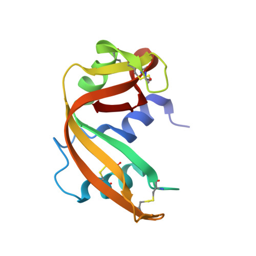
Ionic interactions in crystalline bovine pancreatic ribonuclease A.
Fedorov, A.A., Joseph-McCarthy, D., Fedorov, E., Sirakova, D., Graf, I., Almo, S.C.(1996) Biochemistry 35: 15962-15979
- PubMed: 8973167
- DOI: https://doi.org/10.1021/bi961533g
- Primary Citation of Related Structures:
1RNO, 1RNQ, 1RNW, 1RNX, 1RNY, 1RNZ - PubMed Abstract:
Isomorphous crystals (space group P3(2)21) of bovine pancreatic ribonuclease A (RNase A) were prepared at a pH of 5.5 in a series of high salt conditions, where both the nature of the ions and the ionic strength varied: 80% ammonium sulfate (mu = 12.5); 8 M sodium formate (mu = 8.0); 3 M NaCl, 30% ammonium sulfate (mu = 7.0); 3 M CsCl, 30% ammonium sulfate (mu = 7.0); and 2.5 M NaCl, 3.3 M sodium formate (mu = 5.8). These structures were independently refined to a resolution of 2.0 A or better with R-factors that range from 16.1% to 17.5%. A comparison of these six structures and the monoclinic crystal form of RNase A grown from alcohol shows that changes in ionic strength do not alter the secondary or tertiary structure and that there are no significant changes in intramolecular salt bridges. These findings support the notion that structures determined from crystals grown in high salt are representative of the overall structural and electrostatic features present under physiological conditions. While little effect was observed on the main chain conformation, several residues adopted different side chain conformations and altered hydrogen-bonding patterns, either as result of direct anion binding or more subtle indirect effects. Changes in the ionic composition of the mother liquor allowed for the occupancy of the active site with different anions. The direct observation of active site-bound chloride and formate anions supports the proposal that these species act as true competitive inhibitors of RNase A and not through nonspecific electrostatic effects. The identification of bound formate anions allowed for an experimental validation of computational-based functional group mapping techniques and suggests a useful modification to these approaches. Electrostatic surface potential calculations identify a nearly continuous band of positive potential, consistent with an extended binding site for polynucleotide ligands and substrates. The majority of these residues are not involved in salt bridges, which may facilitate binding to extended polynucleotide substrates. Selection of the appropriate solvent conditions results in an unoccupied active site, which will allow this crystal form to be used for the crystallographic study of productive ligand-binding modes.
Organizational Affiliation:
Department of Biochemistry, Albert Einstein College of Medicine, Bronx, New York 10461, USA.


