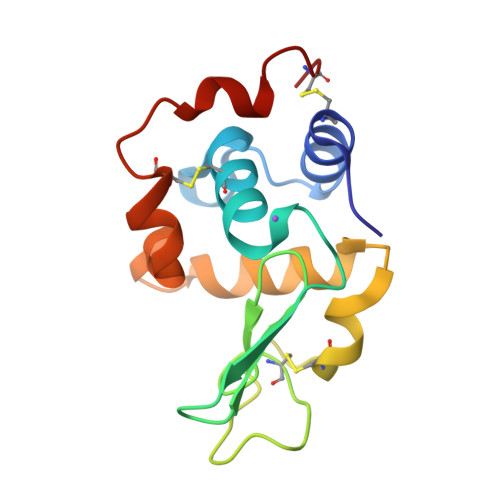
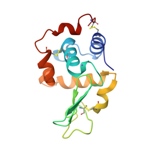
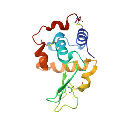
1IX0

I59A-3SS human lysozyme


Experimental Data Snapshot

  • Method: X-RAY DIFFRACTION
  • Resolution: 1.80 Å
  • R-Value Work: 0.179 

wwPDB Validation   3D Report Full Report


This is version 1.5 of the entry. See complete history


Literature

Buried water molecules contribute to the conformational stability of a protein

Takano, K.Yamagata, Y.Yutani, K.

(2003) Protein Eng 16: 5-9

  • DOI: https://doi.org/10.1093/proeng/gzg001
  • Primary Citation of Related Structures:  
    1IX0

  • PubMed Abstract: 

    This study sought to attain a better understanding of the contribution of buried water molecules to protein stability. The 3SS human lysozyme lacks one disulfide bond between Cys77 and Cys95 and is significantly destabilized compared with the wild-type human lysozyme (4SS). We examined the structure and stability of the I59A-3SS mutant human lysozyme, in which a cavity is created at the mutation site. The crystal structure of I59A-3SS indicated that there were ordered new water molecules in the cavity created. The stability of I59A-3SS is 5.5 kJ/mol less than that of 3SS. The decreased stability of I59A-3SS (5.5 kJ/mol) is similar to that of Ile to Ala mutants with newly introduced water molecules in other globular proteins (6.3 +/- 2.1 kJ/mol), but is less than that of Ile/Leu to Ala mutants with empty cavities (13.7 +/- 3.1 kJ/mol). This indicates that water molecules partially compensate for the destabilization by decreasing hydrophobic and van der Waals interactions. These results provide further evidence that buried water molecules contribute to protein stability.


  • Organizational Affiliation

    Institute for Protein Research, Osaka University, Yamadaoka, Suita, Osaka 565-0871, Japan. ktakano@mls.eng.osaka-u.ac.jp


Macromolecules
Find similar proteins by:  (by identity cutoff)  |  3D Structure
Entity ID: 1
MoleculeChains Sequence LengthOrganismDetailsImage
lysozyme130Homo sapiensMutation(s): 3 
EC: 3.2.1.17
UniProt & NIH Common Fund Data Resources
Find proteins for P61626 (Homo sapiens)
Explore P61626 
Go to UniProtKB:  P61626
PHAROS:  P61626
GTEx:  ENSG00000090382 
Entity Groups  
Sequence Clusters30% Identity50% Identity70% Identity90% Identity95% Identity100% Identity
UniProt GroupP61626
Sequence Annotations
Expand
  • Reference Sequence
Small Molecules
Ligands 1 Unique
IDChains Name / Formula / InChI Key2D Diagram3D Interactions
NA
Query on NA

Download Ideal Coordinates CCD File 
B [auth A]SODIUM ION
Na
FKNQFGJONOIPTF-UHFFFAOYSA-N
Experimental Data & Validation

Experimental Data

  • Method: X-RAY DIFFRACTION
  • Resolution: 1.80 Å
  • R-Value Work: 0.179 
  • Space Group: P 21 21 21
Unit Cell:
Length ( Å )Angle ( ˚ )
a = 56.83α = 90
b = 60.88β = 90
c = 32.1γ = 90
Software Package:
Software NamePurpose
DENZOdata reduction
SCALEPACKdata scaling
X-PLORrefinement

Structure Validation

View Full Validation Report


Entry History 

Deposition Data

Revision History  (Full details and data files)

  • Version 1.0: 2003-07-22
    Type: Initial release
  • Version 1.1: 2008-04-27
    Changes: Version format compliance
  • Version 1.2: 2011-07-13
    Changes: Version format compliance
  • Version 1.3: 2021-11-10
    Changes: Database references, Derived calculations
  • Version 1.4: 2023-12-27
    Changes: Data collection
  • Version 1.5: 2024-10-09
    Changes: Structure summary