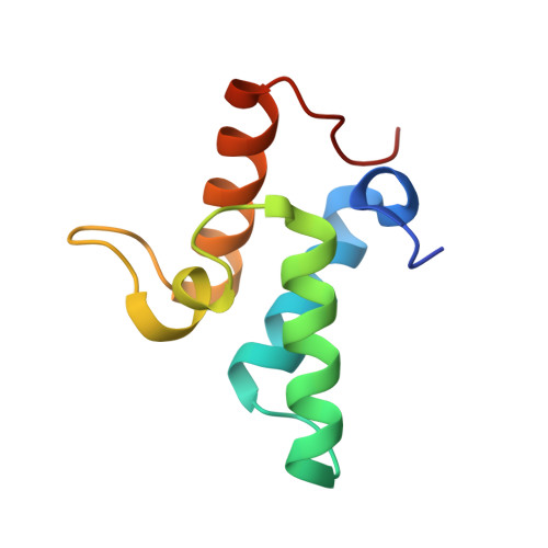
Specificity in protein-protein interactions: the structural basis for dual recognition in endonuclease colicin-immunity protein complexes.
Kuhlmann, U.C., Pommer, A.J., Moore, G.R., James, R., Kleanthous, C.(2000) J Mol Biol 301: 1163-1178
- PubMed: 10966813
- DOI: https://doi.org/10.1006/jmbi.2000.3945
- Primary Citation of Related Structures:
1EMV - PubMed Abstract:
Bacteria producing endonuclease colicins are protected against their cytotoxic activity by virtue of a small immunity protein that binds with high affinity and specificity to inactivate the endonuclease. DNase binding by the immunity protein occurs through a "dual recognition" mechanism in which conserved residues from helix III act as the binding-site anchor, while variable residues from helix II define specificity. We now report the 1.7 A crystal structure of the 24.5 kDa complex formed between the endonuclease domain of colicin E9 and its cognate immunity protein Im9, which provides a molecular rationale for this mechanism. Conserved residues of Im9 form a binding-energy hotspot through a combination of backbone hydrogen bonds to the endonuclease, many via buried solvent molecules, and hydrophobic interactions at the core of the interface, while the specificity-determining residues interact with corresponding specificity side-chains on the enzyme. Comparison between the present structure and that reported recently for the colicin E7 endonuclease domain in complex with Im7 highlights how specificity is achieved by very different interactions in the two complexes, predominantly hydrophobic in nature in the E9-Im9 complex but charged in the E7-Im7 complex. A key feature of both complexes is the contact between a conserved tyrosine residue from the immunity proteins (Im9 Tyr54) with a specificity residue on the endonuclease directing it toward the specificity sites of the immunity protein. Remarkably, this tyrosine residue and its neighbour (Im9 Tyr55) are the pivots of a 19 degrees rigid-body rotation that relates the positions of Im7 and Im9 in the two complexes. This rotation does not affect conserved immunity protein interactions with the endonuclease but results in different regions of the specificity helix being presented to the enzyme.
Organizational Affiliation:
School of Biological Sciences, University of East Anglia, Norwich NR4 7TJ, UK.


