Structure of a transiently phosphorylated switch in bacterial signal transduction.
Kern, D., Volkman, B.F., Luginbuhl, P., Nohaile, M.J., Kustu, S., Wemmer, D.E.(1999) Nature 402: 894-898
- PubMed: 10622255
- DOI: https://doi.org/10.1038/47273
- Primary Citation of Related Structures:
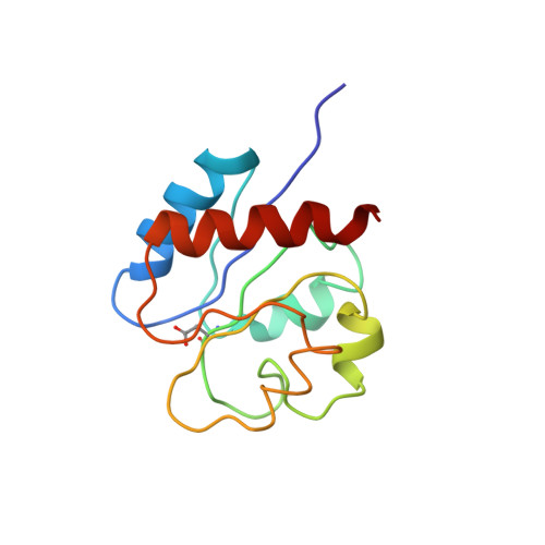
1DC7, 1DC8 - PubMed Abstract:
Receiver domains are the dominant molecular switches in bacterial signalling. Although several structures of non-phosphorylated receiver domains have been reported, a detailed structural understanding of the activation arising from phosphorylation has been impeded by the very short half-lives of the aspartylphosphate linkages. Here we present the first structure of a receiver domain in its active state, the phosphorylated receiver domain of the bacterial enhancer-binding protein NtrC (nitrogen regulatory protein C). Nuclear magnetic resonance spectra were taken during steady-state autophosphorylation/dephosphorylation, and three-dimensional spectra from multiple samples were combined. Phosphorylation induces a large conformational change involving a displacement of beta-strands 4 and 5 and alpha-helices 3 and 4 away from the active site, a register shift and an axial rotation in helix 4. This creates an exposed hydrophobic surface that is likely to transmit the signal to the transcriptional activation domain.
Organizational Affiliation:
Department of Biochemistry, Brandeis University, Waltham, Massachusetts 02454, USA. dkern@brandeis.edu