6TTK

Crystal structure of the kelch domain of human KLHL12 in complex with DVL1 peptide


Experimental Data Snapshot

  • Method: X-RAY DIFFRACTION
  • Resolution: 2.38 Å
  • R-Value Free: 0.251 
  • R-Value Work: 0.225 
  • R-Value Observed: 0.227 

wwPDB Validation   3D Report Full Report


This is version 1.2 of the entry. See complete history


Literature

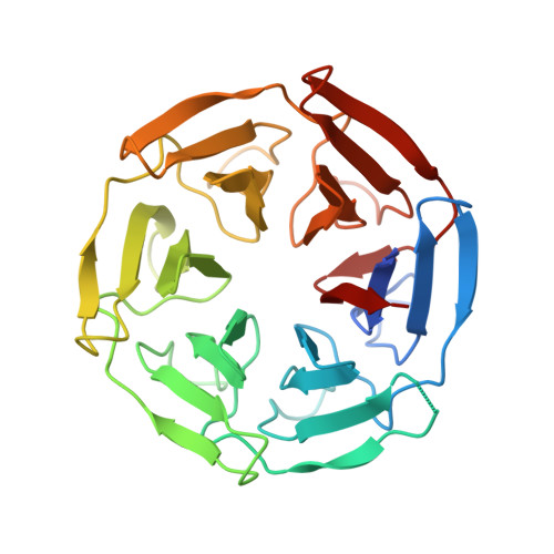
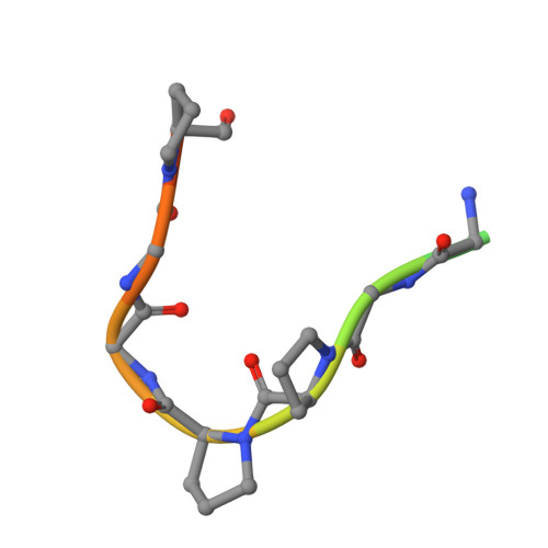
Identification of a PGXPP degron motif in dishevelled and structural basis for its binding to the E3 ligase KLHL12.

Chen, Z.Wasney, G.A.Picaud, S.Filippakopoulos, P.Vedadi, M.D'Angiolella, V.Bullock, A.N.

(2020) Open Biol 10: 200041-200041

  • DOI: https://doi.org/10.1098/rsob.200041
  • Primary Citation of Related Structures:  
    6TTK

  • PubMed Abstract: 

    Wnt signalling is dependent on dishevelled proteins (DVL1-3), which assemble an intracellular Wnt signalosome at the plasma membrane. The levels of DVL1-3 are regulated by multiple Cullin-RING E3 ligases that mediate their ubiquitination and degradation. The BTB-Kelch protein KLHL12 was the first E3 ubiquitin ligase to be identified for DVL1-3, but the molecular mechanisms determining its substrate interactions have remained unknown. Here, we mapped the interaction of DVL1-3 to a 'PGXPP' motif that is conserved in other known partners and substrates of KLHL12, including PLEKHA4, PEF1, SEC31 and DRD4. To determine the binding mechanism, we solved a 2.4 Å crystal structure of the Kelch domain of KLHL12 in complex with a DVL1 peptide that bound with low micromolar affinity. The DVL1 substrate adopted a U-shaped turn conformation that enabled hydrophobic interactions with all six blades of the Kelch domain β-propeller. In cells, the mutation or deletion of this motif reduced the binding and ubiquitination of DVL1 and increased its stability confirming this sequence as a degron motif for KLHL12 recruitment. These results define the molecular mechanisms determining DVL regulation by KLHL12 and establish the KLHL12 Kelch domain as a new protein interaction module for a novel proline-rich motif.


  • Organizational Affiliation

    Structural Genomics Consortium, University of Oxford, Old Road Campus Research Building, Roosevelt Drive, Oxford OX3 7DQ, UK.


Macromolecules
Find similar proteins by:  (by identity cutoff)  |  3D Structure
Entity ID: 1
MoleculeChains Sequence LengthOrganismDetailsImage
Kelch-like protein 12
A, B, C, D
301Homo sapiensMutation(s): 0 
Gene Names: KLHL12C3IP1
UniProt & NIH Common Fund Data Resources
Find proteins for Q53G59 (Homo sapiens)
Explore Q53G59 
Go to UniProtKB:  Q53G59
PHAROS:  Q53G59
GTEx:  ENSG00000117153 
Entity Groups  
Sequence Clusters30% Identity50% Identity70% Identity90% Identity95% Identity100% Identity
UniProt GroupQ53G59
Sequence Annotations
Expand
  • Reference Sequence

Find similar proteins by:  Sequence   |   3D Structure  

Entity ID: 2
MoleculeChains Sequence LengthOrganismDetailsImage
DVL1E [auth F],
F [auth G],
G [auth H],
H [auth E]
15Homo sapiensMutation(s): 0 
UniProt & NIH Common Fund Data Resources
Find proteins for O14640 (Homo sapiens)
Explore O14640 
Go to UniProtKB:  O14640
PHAROS:  O14640
GTEx:  ENSG00000107404 
Entity Groups  
Sequence Clusters30% Identity50% Identity70% Identity90% Identity95% Identity100% Identity
UniProt GroupO14640
Sequence Annotations
Expand
  • Reference Sequence
Experimental Data & Validation

Experimental Data

  • Method: X-RAY DIFFRACTION
  • Resolution: 2.38 Å
  • R-Value Free: 0.251 
  • R-Value Work: 0.225 
  • R-Value Observed: 0.227 
  • Space Group: P 1 21 1
Unit Cell:
Length ( Å )Angle ( ˚ )
a = 80.225α = 90
b = 73.145β = 94.5
c = 101.845γ = 90
Software Package:
Software NamePurpose
PHENIXrefinement
Aimlessdata scaling
PHASERphasing
PDB_EXTRACTdata extraction
xia2data reduction

Structure Validation

View Full Validation Report


Entry History 

Revision History  (Full details and data files)

  • Version 1.0: 2020-02-12
    Type: Initial release
  • Version 1.1: 2020-08-26
    Changes: Database references
  • Version 1.2: 2024-01-24
    Changes: Data collection, Database references, Refinement description