6NJ0

Wild-type E. coli MenE with bound m phenylether-linked analogue of OSB-AMS


Experimental Data Snapshot

  • Method: X-RAY DIFFRACTION
  • Resolution: 1.83 Å
  • R-Value Free: 0.244 
  • R-Value Work: 0.196 
  • R-Value Observed: 0.199 

wwPDB Validation   3D Report Full Report


Ligand Structure Quality Assessment 


This is version 1.2 of the entry. See complete history


Literature

Structure-Based Design, Synthesis, and Biological Evaluation of Non-Acyl Sulfamate Inhibitors of the Adenylate-Forming Enzyme MenE.

Evans, C.E.Si, Y.Matarlo, J.S.Yin, Y.French, J.B.Tonge, P.J.Tan, D.S.

(2019) Biochemistry 58: 1918-1930

  • DOI: https://doi.org/10.1021/acs.biochem.9b00003
  • Primary Citation of Related Structures:  
    6NJ0

  • PubMed Abstract: 

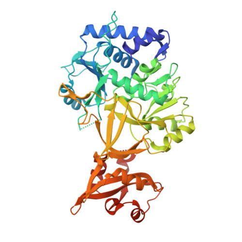
    N-Acyl sulfamoyladenosines (acyl-AMS) have been used extensively to inhibit adenylate-forming enzymes that are involved in a wide range of biological processes. These acyl-AMS inhibitors are nonhydrolyzable mimics of the cognate acyl adenylate intermediates that are bound tightly by adenylate-forming enzymes. However, the anionic acyl sulfamate moiety presents a pharmacological liability that may be detrimental to cell permeability and pharmacokinetic profiles. We have previously developed the acyl sulfamate OSB-AMS (1) as a potent inhibitor of the adenylate-forming enzyme MenE, an o-succinylbenzoate-CoA (OSB-CoA) synthetase that is required for bacterial menaquinone biosynthesis. Herein, we report the use of computational docking to develop novel, non-acyl sulfamate inhibitors of MenE. A m-phenyl ether-linked analogue (5) was found to be the most potent inhibitor (IC 50 = 8 μM; K d = 244 nM), and its X-ray co-crystal structure was determined to characterize its binding mode in comparison to the computational prediction. This work provides a framework for the development of potent non-acyl sulfamate inhibitors of other adenylate-forming enzymes in the future.


Macromolecules
Find similar proteins by:  (by identity cutoff)  |  3D Structure
Entity ID: 1
MoleculeChains Sequence LengthOrganismDetailsImage
2-succinylbenzoate--CoA ligase451Escherichia coli K-12Mutation(s): 0 
Gene Names: menEb2260JW2255
EC: 6.2.1.26
UniProt
Find proteins for P37353 (Escherichia coli (strain K12))
Explore P37353 
Go to UniProtKB:  P37353
Entity Groups  
Sequence Clusters30% Identity50% Identity70% Identity90% Identity95% Identity100% Identity
UniProt GroupP37353
Sequence Annotations
Expand
  • Reference Sequence
Small Molecules
Ligands 1 Unique
IDChains Name / Formula / InChI Key2D Diagram3D Interactions
KOY
Query on KOY

Download Ideal Coordinates CCD File 
B [auth A]5'-O-{3-[3-(2-carboxyphenyl)-3-oxopropyl]phenyl}adenosine
C26 H25 N5 O7
GEMGRSMVWNUQGG-PTGPVQHPSA-N
Binding Affinity Annotations 
IDSourceBinding Affinity
KOY Binding MOAD:  6NJ0 IC50: 8100 (nM) from 1 assay(s)
Experimental Data & Validation

Experimental Data

  • Method: X-RAY DIFFRACTION
  • Resolution: 1.83 Å
  • R-Value Free: 0.244 
  • R-Value Work: 0.196 
  • R-Value Observed: 0.199 
  • Space Group: P 21 21 21
Unit Cell:
Length ( Å )Angle ( ˚ )
a = 71.146α = 90
b = 74.424β = 90
c = 79.033γ = 90
Software Package:
Software NamePurpose
REFMACrefinement
XDSdata reduction
Aimlessdata scaling
MOLREPphasing

Structure Validation

View Full Validation Report


Ligand Structure Quality Assessment 


Entry History 

Deposition Data

Revision History  (Full details and data files)

  • Version 1.0: 2019-04-10
    Type: Initial release
  • Version 1.1: 2019-04-17
    Changes: Data collection, Database references
  • Version 1.2: 2024-03-13
    Changes: Data collection, Database references