6HE2 | pdb_00006he2

Crystal structure of an open conformation of 2-Hydroxyisobutyryl-CoA Ligase (HCL) in complex with 2-HIB-AMP and CoA


Experimental Data Snapshot

  • Method: X-RAY DIFFRACTION
  • Resolution: 2.30 Å
  • R-Value Free: 
    0.222 (Depositor), 0.228 (DCC) 
  • R-Value Work: 
    0.184 (Depositor), 0.186 (DCC) 
  • R-Value Observed: 
    0.186 (Depositor) 

Starting Model: experimental
View more details

wwPDB Validation 3D Report Full Report

Validation slider image for 6HE2

Ligand Structure Quality Assessment 


This is version 1.1 of the entry. See complete history

Literature

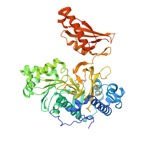
Structures of 2-Hydroxyisobutyric Acid-CoA Ligase Reveal Determinants of Substrate Specificity and Describe a Multi-Conformational Catalytic Cycle.

Zahn, M.Kurteva-Yaneva, N.Schuster, J.Krug, U.Georgi, T.Muller, R.H.Rohwerder, T.Strater, N.

(2019) J Mol Biology 431: 2747-2761

  • DOI: https://doi.org/10.1016/j.jmb.2019.05.027
  • Primary Citation Related Structures: 
    6HDW, 6HDX, 6HDY, 6HE0, 6HE2

  • PubMed Abstract: 

    2-Hydroxyisobutyric acid (2-HIBA) is a biomarker of adiposity and associated metabolic diseases such as diabetes mellitus. It is also formed in the bacterial degradation pathway of the fuel oxygenate methyl tert-butyl ether (MTBE), requiring thioesterification with CoA prior to isomerization to 3-hydroxybutyryl-CoA by B 12 -dependent acyl-CoA mutases. Here, we identify the adenylating enzymes superfamily member 2-HIBA-CoA ligase (HCL) in the MTBE-degrading bacterium Aquincola tertiaricarbonis L108 by knockout experiments. To characterize this central enzyme of 2-HIBA metabolism, ligase activity kinetics of purified HCL and its X-ray crystal structures were studied. We analyzed the enzyme in three states, which differ in the orientation of the two enzyme domains. A 154° rotation of the C-terminal domain accompanies the switch from the adenylate- into the thioester-forming state. Furthermore, a third conformation was obtained, which differs by 50° and 130° from the adenylation and thioesterification states, respectively. Phylogenetic and structural analysis reveals that HCL defines a new subgroup within phenylacetate-CoA ligases (PCLs) thus far described to exclusively accept aromatic acyl substrates. In contrast, kinetic characterization clearly demonstrated that HCL catalyzes CoA activation of several aliphatic short-chain carboxylic acids, preferentially 2-HIBA. Compared to the classical PCL representatives PaaK1 and PaaK2 of Burkholderia cenocepacia J2315, the acyl binding pocket of HCL is significantly smaller and more polar, due to unique active-site residues Y164 and S239 forming H-bonds with the OH-group of the acyl substrate moiety. Furthermore, HCL and PaaK topologies illustrate the evolutionary steps leading from a homodimeric to the fused monomeric core fold found in other ligases.


  • Organizational Affiliation
    • Institute of Bioanalytical Chemistry, Center for Biotechnology and Biomedicine, Leipzig University, Deutscher Platz 5, 04103 Leipzig, Germany.

Macromolecule Content 

  • Total Structure Weight: 114.98 kDa 
  • Atom Count: 7,501 
  • Modeled Residue Count: 890 
  • Deposited Residue Count: 998 
  • Unique protein chains: 1

Macromolecules

Find similar proteins by:|  3D Structure
Entity ID: 1
MoleculeChains  Sequence LengthOrganismDetailsImage
2-hydroxyisobutyryl-CoA synthetase
A, B
499Aquincola tertiaricarbonisMutation(s): 0 
Gene Names: hcl
EC: 6.2.1
UniProt
Find proteins for I3VE75 (Aquincola tertiaricarbonis)
Explore I3VE75 
Go to UniProtKB:  I3VE75
Entity Groups
Sequence Clusters30% Identity50% Identity70% Identity90% Identity95% Identity100% Identity
UniProt GroupI3VE75
Sequence Annotations
Expand
Reference Sequence

Small Molecules

Ligands 3 Unique
IDChains Name / Formula / InChI Key2D Diagram3D Interactions
COA

Query on COA


Download:Ideal Coordinates CCD File
E [auth A],
H [auth B]
COENZYME A
C21 H36 N7 O16 P3 S
RGJOEKWQDUBAIZ-IBOSZNHHSA-N
8LE

Query on 8LE


Download:Ideal Coordinates CCD File
D [auth A],
G [auth B]
[[(2~{R},3~{S},4~{R},5~{R})-5-(6-aminopurin-9-yl)-3,4-bis(oxidanyl)oxolan-2-yl]methoxy-oxidanyl-phosphoryl] 2-methyl-2-oxidanyl-propanoate
C14 H20 N5 O9 P
WJCYMTFWIDDQPQ-WOUKDFQISA-N
AMP

Query on AMP


Download:Ideal Coordinates CCD File
C [auth A],
F [auth B]
ADENOSINE MONOPHOSPHATE
C10 H14 N5 O7 P
UDMBCSSLTHHNCD-KQYNXXCUSA-N

Experimental Data & Validation

Experimental Data

  • Method: X-RAY DIFFRACTION
  • Resolution: 2.30 Å
  • R-Value Free:  0.222 (Depositor), 0.228 (DCC) 
  • R-Value Work:  0.184 (Depositor), 0.186 (DCC) 
  • R-Value Observed: 0.186 (Depositor) 
Space Group: P 21 21 21
Unit Cell:
Length ( Å )Angle ( ˚ )
a = 93.852α = 90
b = 101.847β = 90
c = 109.48γ = 90
Software Package:
Software NamePurpose
XDSdata reduction
Aimlessdata scaling
MOLREPphasing
BUSTERrefinement

Structure Validation

View Full Validation Report


Ligand Structure Quality Assessment 


Entry History 

Deposition Data

Revision History  (Full details and data files)

  • Version 1.0: 2019-08-28
    Type: Initial release
  • Version 1.1: 2024-01-17
    Changes: Advisory, Data collection, Database references, Refinement description