6H53

Crystal structure of Mycobacterium tuberculosis phosphatidylinositol phosphate synthase (PgsA1) in apo form


Experimental Data Snapshot

  • Method: X-RAY DIFFRACTION
  • Resolution: 2.90 Å
  • R-Value Free: 0.261 
  • R-Value Work: 0.220 
  • R-Value Observed: 0.222 

wwPDB Validation   3D Report Full Report


Ligand Structure Quality Assessment 


This is version 1.2 of the entry. See complete history


Literature

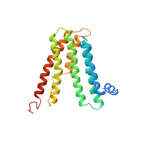
Structure ofMycobacterium tuberculosisphosphatidylinositol phosphate synthase reveals mechanism of substrate binding and metal catalysis.

Grave, K.Bennett, M.D.Hogbom, M.

(2019) Commun Biol 2: 175-175

  • DOI: https://doi.org/10.1038/s42003-019-0427-1
  • Primary Citation of Related Structures:  
    6H53, 6H59, 6H5A

  • PubMed Abstract: 

    Tuberculosis causes over one million yearly deaths, and drug resistance is rapidly developing. Mycobacterium tuberculosis phosphatidylinositol phosphate synthase (PgsA1) is an integral membrane enzyme involved in biosynthesis of inositol-derived phospholipids required for formation of the mycobacterial cell wall, and a potential drug target. Here we present three crystal structures of M. tuberculosis PgsA1: in absence of substrates (2.9 Å), in complex with Mn 2+ and citrate (1.9 Å), and with the CDP-DAG substrate (1.8 Å). The structures reveal atomic details of substrate binding as well as coordination and dynamics of the catalytic metal site. In addition, molecular docking supported by mutagenesis indicate a binding mode for the second substrate, D- myo -inositol-3-phosphate. Together, the data describe the structural basis for M. tuberculosis phosphatidylinositol phosphate synthesis and suggest a refined general catalytic mechanism-including a substrate-induced carboxylate shift-for Class I CDP-alcohol phosphotransferases, enzymes essential for phospholipid biosynthesis in all domains of life.


  • Organizational Affiliation

    Department of Biochemistry and Biophysics, Stockholm University, Svante Arrhenius väg 16 C, SE-106 91 Stockholm, Sweden.


Macromolecules
Find similar proteins by:  (by identity cutoff)  |  3D Structure
Entity ID: 1
MoleculeChains Sequence LengthOrganismDetailsImage
CDP-diacylglycerol--inositol 3-phosphatidyltransferase
A, B
217Mycobacterium tuberculosis H37RvMutation(s): 0 
Gene Names: pgsA1Rv2612c
EC: 2.7.8.11
Membrane Entity: Yes 
UniProt
Find proteins for P9WPG7 (Mycobacterium tuberculosis (strain ATCC 25618 / H37Rv))
Explore P9WPG7 
Go to UniProtKB:  P9WPG7
Entity Groups  
Sequence Clusters30% Identity50% Identity70% Identity90% Identity95% Identity100% Identity
UniProt GroupP9WPG7
Sequence Annotations
Expand
  • Reference Sequence
Small Molecules
Ligands 2 Unique
IDChains Name / Formula / InChI Key2D Diagram3D Interactions
UPL
Query on UPL

Download Ideal Coordinates CCD File 
D [auth A]
E [auth A]
F [auth A]
G [auth A]
J [auth B]
D [auth A],
E [auth A],
F [auth A],
G [auth A],
J [auth B],
K [auth B]
UNKNOWN BRANCHED FRAGMENT OF PHOSPHOLIPID
C34 H70
QPCSNFPFMCVVAI-UHFFFAOYSA-N
SO4
Query on SO4

Download Ideal Coordinates CCD File 
C [auth A],
H [auth A],
I [auth B]
SULFATE ION
O4 S
QAOWNCQODCNURD-UHFFFAOYSA-L
Experimental Data & Validation

Experimental Data

  • Method: X-RAY DIFFRACTION
  • Resolution: 2.90 Å
  • R-Value Free: 0.261 
  • R-Value Work: 0.220 
  • R-Value Observed: 0.222 
  • Space Group: P 21 21 21
Unit Cell:
Length ( Å )Angle ( ˚ )
a = 69.726α = 90
b = 72.896β = 90
c = 102.452γ = 90
Software Package:
Software NamePurpose
SCALAdata scaling
PHASERphasing
PHENIXrefinement
PDB_EXTRACTdata extraction
XDSdata reduction

Structure Validation

View Full Validation Report


Ligand Structure Quality Assessment 


Entry History 

Deposition Data

Revision History  (Full details and data files)

  • Version 1.0: 2019-05-15
    Type: Initial release
  • Version 1.1: 2019-05-29
    Changes: Data collection, Database references
  • Version 1.2: 2024-01-17
    Changes: Data collection, Database references, Refinement description