6M75

C-Myc DNA binding protein complex


Experimental Data Snapshot

  • Method: X-RAY DIFFRACTION
  • Resolution: 2.57 Å
  • R-Value Free: 0.236 
  • R-Value Work: 0.211 
  • R-Value Observed: 0.214 

wwPDB Validation   3D Report Full Report


This is version 2.1 of the entry. See complete history


Literature

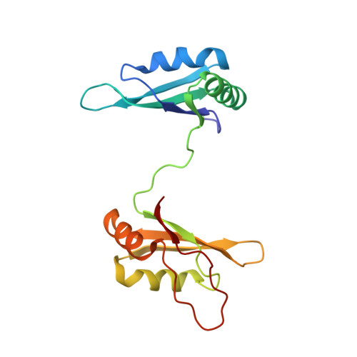
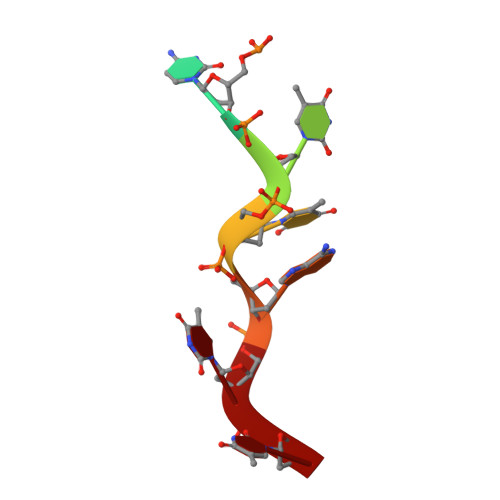
Hinge like domain motion facilitates human RBMS1 protein binding to proto-oncogene c-myc promoter.

Aggarwal, P.Bhavesh, N.S.

(2021) Nucleic Acids Res 49: 5943-5955

  • DOI: https://doi.org/10.1093/nar/gkab363
  • Primary Citation of Related Structures:  
    6M75, 7C36

  • PubMed Abstract: 

    DNA binding proteins recognize DNA specifically or non-specifically using direct and indirect readout mechanisms like sliding, hopping, and diffusion. However, a common difficulty in explicitly elucidating any particular mechanism of site-specific DNA-protein recognition is the lack of knowledge regarding target sequences and inadequate account of non-specific interactions, in general. Here, we decipher the structural basis of target search performed by the key regulator of expression of c-myc proto-oncogene, the human RBMS1 protein. In this study, we have shown the structural reorganization of this multi-domain protein required for recognizing the specific c-myc promoter sequence. The results suggest that a synergy between structural re-organization and thermodynamics is necessary for the recognition of target sequences. The study presents another perspective of looking at the DNA-protein interactions.


  • Organizational Affiliation

    Transcription Regulation group, International Centre for Genetic Engineering and Biotechnology (ICGEB), Aruna Asaf Ali Marg, New Delhi 110067, India.


Macromolecules

Find similar proteins by:  (by identity cutoff)  |  3D Structure
Entity ID: 1
MoleculeChains Sequence LengthOrganismDetailsImage
RNA-binding motif, single-stranded-interacting protein 1167Homo sapiensMutation(s): 0 
Gene Names: RBMS1C2orf12MSSPMSSP1SCR2
UniProt & NIH Common Fund Data Resources
Find proteins for P29558 (Homo sapiens)
Explore P29558 
Go to UniProtKB:  P29558
PHAROS:  P29558
GTEx:  ENSG00000153250 
Entity Groups  
Sequence Clusters30% Identity50% Identity70% Identity90% Identity95% Identity100% Identity
UniProt GroupP29558
Sequence Annotations
Expand
  • Reference Sequence

Find similar nucleic acids by:  Sequence   |   3D Structure  

Entity ID: 2
MoleculeChains LengthOrganismImage
DNA (5'-D(*TP*CP*TP*TP*AP*TP*T)-3')B [auth C]7Homo sapiens
Sequence Annotations
Expand
  • Reference Sequence
Experimental Data & Validation

Experimental Data

Unit Cell:
Length ( Å )Angle ( ˚ )
a = 83.68α = 90
b = 114.946β = 90
c = 27.426γ = 90
Software Package:
Software NamePurpose
PHENIXrefinement
XDSdata reduction
autoPROCdata scaling
Auto-Rickshawphasing

Structure Validation

View Full Validation Report


Entry History 

Deposition Data

Revision History  (Full details and data files)

  • Version 1.0: 2021-03-17
    Type: Initial release
  • Version 1.1: 2021-07-14
    Changes: Database references
  • Version 2.0: 2022-06-22
    Changes: Database references, Polymer sequence
  • Version 2.1: 2023-11-29
    Changes: Data collection, Refinement description