The crystal structure of nitroreductase A from Vibrio parahaemolyticus RIMD 2210633
Tan, K., Zhou, M., Anderson, W.F., Joachimiak, A.To be published.
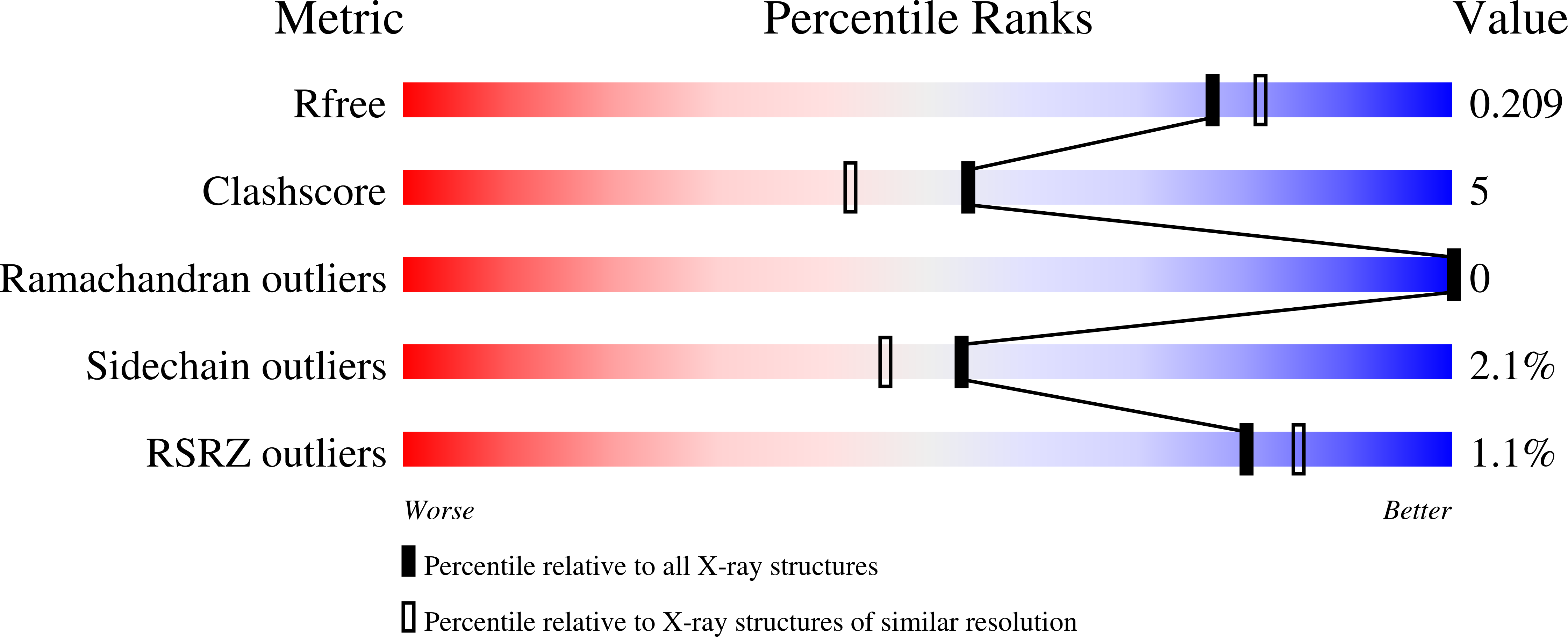
Experimental Data Snapshot
Starting Model: experimental
View more details
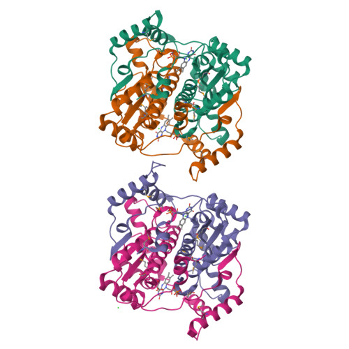
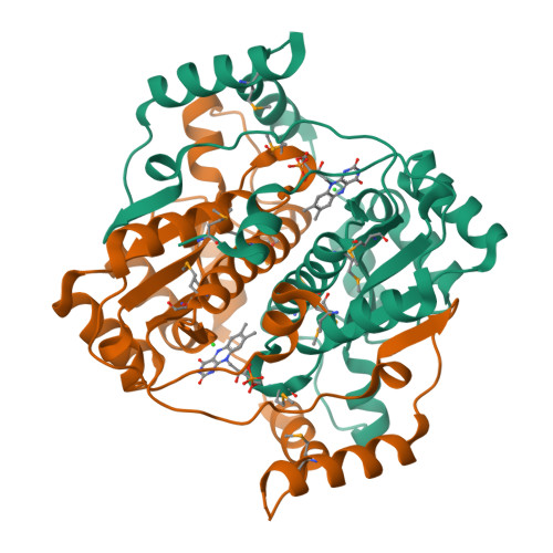
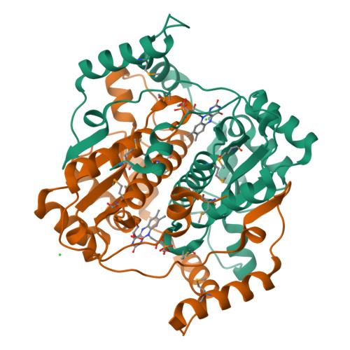
Entity ID: 1 | |||||
|---|---|---|---|---|---|
| Molecule | Chains | Sequence Length | Organism | Details | Image |
| NADPH-flavin oxidoreductase | 243 | Vibrio parahaemolyticus RIMD 2210633 | Mutation(s): 0 Gene Names: VPA1601 | 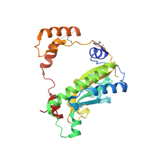 | |
UniProt | |||||
Find proteins for Q87FS7 (Vibrio parahaemolyticus serotype O3:K6 (strain RIMD 2210633)) Explore Q87FS7 Go to UniProtKB: Q87FS7 | |||||
Entity Groups | |||||
| Sequence Clusters | 30% Identity50% Identity70% Identity90% Identity95% Identity100% Identity | ||||
| UniProt Group | Q87FS7 | ||||
Sequence AnnotationsExpand | |||||
| |||||
| Ligands 3 Unique | |||||
|---|---|---|---|---|---|
| ID | Chains | Name / Formula / InChI Key | 2D Diagram | 3D Interactions | |
| FMN Query on FMN | E [auth A], G [auth B], I [auth C], K [auth D] | FLAVIN MONONUCLEOTIDE C17 H21 N4 O9 P FVTCRASFADXXNN-SCRDCRAPSA-N |  | ||
| GOL Query on GOL | L [auth D] | GLYCEROL C3 H8 O3 PEDCQBHIVMGVHV-UHFFFAOYSA-N |  | ||
| CL Query on CL | F [auth A], H [auth B], J [auth C], M [auth D] | CHLORIDE ION Cl VEXZGXHMUGYJMC-UHFFFAOYSA-M |  | ||
| Modified Residues 1 Unique | |||||
|---|---|---|---|---|---|
| ID | Chains | Type | Formula | 2D Diagram | Parent |
| MSE Query on MSE | A, B, C, D | L-PEPTIDE LINKING | C5 H11 N O2 Se |  | MET |
| Length ( Å ) | Angle ( ˚ ) |
|---|---|
| a = 57.004 | α = 90 |
| b = 71.609 | β = 93.15 |
| c = 110.45 | γ = 90 |
| Software Name | Purpose |
|---|---|
| PHENIX | refinement |
| HKL-3000 | data reduction |
| HKL-3000 | data scaling |
| HKL-3000 | phasing |
| Funding Organization | Location | Grant Number |
|---|---|---|
| National Institutes of Health/National Institute Of Allergy and Infectious Diseases (NIH/NIAID) | United States | HHSN272201200026C |
RCSB PDB is hosted by

RCSB PDB is a member of the