5U5G | pdb_00005u5g

Psf3 in complex with NADP+ and 2-OPP


Experimental Data Snapshot

  • Method: X-RAY DIFFRACTION
  • Resolution: 2.05 Å
  • R-Value Free: 
    0.207 (Depositor), 0.180 (DCC) 
  • R-Value Work: 
    0.170 (Depositor), 0.170 (DCC) 
  • R-Value Observed: 
    0.172 (Depositor) 

wwPDB Validation   3D Report Full Report


Ligand Structure Quality Assessment 

Created with Raphaël 2.3.0Worse 01 BetterLigand structure goodness of fit to experimental dataBest fitted NAPClick on this verticalbar to view details

This is version 1.5 of the entry. See complete history


Literature

Characterization of Two Late-Stage Enzymes Involved in Fosfomycin Biosynthesis in Pseudomonads.

Olivares, P.Ulrich, E.C.Chekan, J.R.van der Donk, W.A.Nair, S.K.

(2017) ACS Chem Biol 12: 456-463

  • DOI: https://doi.org/10.1021/acschembio.6b00939
  • Primary Citation of Related Structures:  
    5U55, 5U57, 5U58, 5U5D, 5U5G

  • PubMed Abstract: 

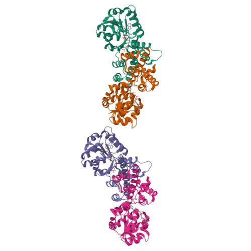
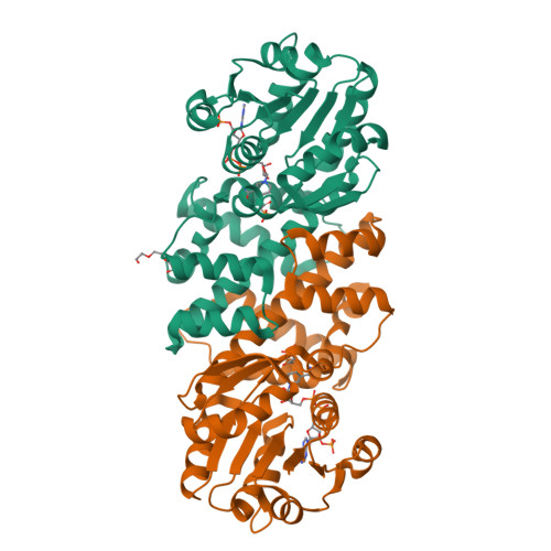
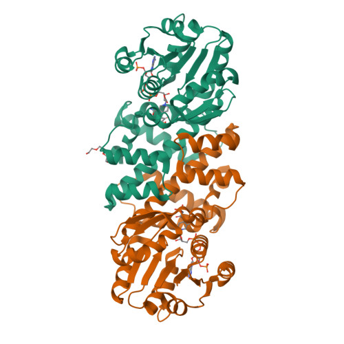
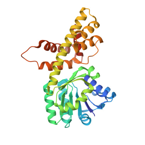
    The broad-spectrum phosphonate antibiotic fosfomycin is currently in use for clinical treatment of infections caused by both Gram-positive and Gram-negative uropathogens. The antibiotic is biosynthesized by various streptomycetes, as well as by pseudomonads. Notably, the biosynthetic strategies used by the two genera share only two steps: the first step in which primary metabolite phosphoenolpyruvate (PEP) is converted to phosphonopyruvate (PnPy) and the terminal step in which 2-hydroxypropylphosphonate (2-HPP) is converted to fosfomycin. Otherwise, distinct enzymatic paths are employed. Here, we biochemically confirm the last two steps in the fosfomycin biosynthetic pathway of Pseudomonas syringae PB-5123, showing that Psf3 performs the reduction of 2-oxopropylphosphonate (2-OPP) to (S)-2-HPP, followed by the Psf4-catalyzed epoxidation of (S)-2-HPP to fosfomycin. Psf4 can also accept (R)-2-HPP as a substrate but instead performs an oxidation to make 2-OPP. We show that the combined activities of Psf3 and Psf4 can be used to convert racemic 2-HPP to fosfomycin in an enantioconvergent process. X-ray structures of each enzyme with bound substrates provide insights into the stereospecificity of each conversion. These studies shed light on the reaction mechanisms of the two terminal enzymes in a distinct pathway employed by pseudomonads for the production of a potent antimicrobial agent.


  • Organizational Affiliation

    Department of Biochemistry, ‡Department of Chemistry, §Carl R. Woese Institute for Genomic Biology, ∥Howard Hughes Medical Institute, and ⊥Center for Biophysics and Computational Biology, University of Illinois at Urbana-Champaign , Urbana, Illinois 61801, United States.


Macromolecules
Find similar proteins by:  (by identity cutoff)  |  3D Structure
Entity ID: 1
MoleculeChains Sequence LengthOrganismDetailsImage
6-phosphogluconate dehydrogenase
A, B, C, D
295Pseudomonas syringaeMutation(s): 0 
UniProt
Find proteins for I6R485 (Pseudomonas syringae)
Explore I6R485 
Go to UniProtKB:  I6R485
Entity Groups  
Sequence Clusters30% Identity50% Identity70% Identity90% Identity95% Identity100% Identity
UniProt GroupI6R485
Sequence Annotations
Expand
  • Reference Sequence
Small Molecules
Ligands 3 Unique
IDChains Name / Formula / InChI Key2D Diagram3D Interactions
NAP
Query on NAP

Download Ideal Coordinates CCD File 
E [auth A],
H [auth B],
J [auth C],
M [auth D]
NADP NICOTINAMIDE-ADENINE-DINUCLEOTIDE PHOSPHATE
C21 H28 N7 O17 P3
XJLXINKUBYWONI-NNYOXOHSSA-N
PGE
Query on PGE

Download Ideal Coordinates CCD File 
G [auth A],
L [auth C]
TRIETHYLENE GLYCOL
C6 H14 O4
ZIBGPFATKBEMQZ-UHFFFAOYSA-N
7VD
Query on 7VD

Download Ideal Coordinates CCD File 
F [auth A],
I [auth B],
K [auth C],
N [auth D]
(2-oxopropyl)phosphonic acid
C3 H7 O4 P
DVSHXLWNRCEDCW-UHFFFAOYSA-N
Experimental Data & Validation

Experimental Data

  • Method: X-RAY DIFFRACTION
  • Resolution: 2.05 Å
  • R-Value Free:  0.207 (Depositor), 0.180 (DCC) 
  • R-Value Work:  0.170 (Depositor), 0.170 (DCC) 
  • R-Value Observed: 0.172 (Depositor) 
Space Group: P 1 21 1
Unit Cell:
Length ( Å )Angle ( ˚ )
a = 46.076α = 90
b = 87.369β = 92.04
c = 164.011γ = 90
Software Package:
Software NamePurpose
PHENIXrefinement
HKL-2000data reduction
HKL-2000data scaling
PHASERphasing

Structure Validation

View Full Validation Report


Ligand Structure Quality Assessment 

Created with Raphaël 2.3.0Worse 01 BetterLigand structure goodness of fit to experimental dataBest fitted NAPClick on this verticalbar to view details

Entry History & Funding Information

Deposition Data


Funding OrganizationLocationGrant Number
National Institutes of Health/National Institute of General Medical Sciences (NIH/NIGMS)United StatesPO1 GM077596

Revision History  (Full details and data files)

  • Version 1.0: 2017-01-04
    Type: Initial release
  • Version 1.1: 2017-03-01
    Changes: Database references
  • Version 1.2: 2017-09-20
    Changes: Author supporting evidence
  • Version 1.3: 2017-11-01
    Changes: Author supporting evidence
  • Version 1.4: 2020-01-01
    Changes: Author supporting evidence
  • Version 1.5: 2024-03-06
    Changes: Data collection, Database references