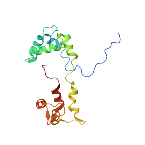
5TBY

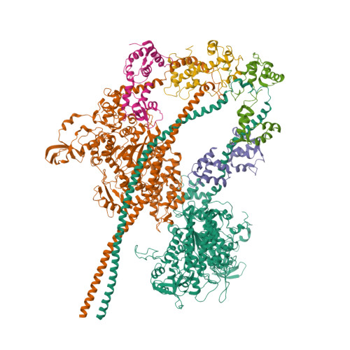
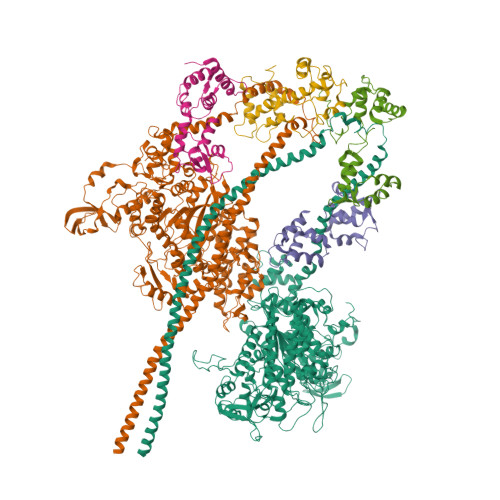
HUMAN BETA CARDIAC HEAVY MEROMYOSIN INTERACTING-HEADS MOTIF OBTAINED BY HOMOLOGY MODELING (USING SWISS-MODEL) OF HUMAN SEQUENCE FROM APHONOPELMA HOMOLOGY MODEL (PDB-3JBH), RIGIDLY FITTED TO HUMAN BETA-CARDIAC NEGATIVELY STAINED THICK FILAMENT 3D-RECONSTRUCTION (EMD-2240)

  • Classification: CONTRACTILE PROTEIN
  • Organism(s): Homo sapiens
  • Mutation(s): No 

  • Deposited: 2016-09-13 Released: 2017-06-07 
  • Deposition Author(s): ALAMO, L., WARE, J.S., PINTO, A., GILLILAN, R.E., SEIDMAN, J.G., SEIDMAN, C.E., PADRON, R.
  • Funding Organization(s): Howard Hughes Medical Institute (HHMI), Leducq Foundation, Wellcome Trust, National Institutes of Health/National Heart, Lung, and Blood Institute (NIH/NHLBI), National Science Foundation (NSF, United States), National Institutes of Health/National Institute of General Medical Sciences (NIH/NIGMS), Medical Research Council (MRC, United Kingdom)

Experimental Data Snapshot

  • Method: ELECTRON MICROSCOPY
  • Resolution: 20.0 Å
  • Aggregation State: FILAMENT 
  • Reconstruction Method: SINGLE PARTICLE 

Starting Model: experimental
View more details

wwPDB Validation   3D Report Full Report


This is version 1.9 of the entry. See complete history


Literature

Effects of myosin variants on interacting-heads motif explain distinct hypertrophic and dilated cardiomyopathy phenotypes.

Alamo, L.Ware, J.S.Pinto, A.Gillilan, R.E.Seidman, J.G.Seidman, C.E.Padron, R.

(2017) Elife 6

  • DOI: https://doi.org/10.7554/eLife.24634
  • Primary Citation of Related Structures:  
    5TBY

  • PubMed Abstract: 

    Cardiac β-myosin variants cause hypertrophic (HCM) or dilated (DCM) cardiomyopathy by disrupting sarcomere contraction and relaxation. The locations of variants on isolated myosin head structures predict contractility effects but not the prominent relaxation and energetic deficits that characterize HCM. During relaxation, pairs of myosins form interacting-heads motif (IHM) structures that with other sarcomere proteins establish an energy-saving, super-relaxed (SRX) state. Using a human β-cardiac myosin IHM quasi-atomic model, we defined interactions sites between adjacent myosin heads and associated protein partners, and then analyzed rare variants from 6112 HCM and 1315 DCM patients and 33,370 ExAC controls. HCM variants, 72% that changed electrostatic charges, disproportionately altered IHM interaction residues (expected 23%; HCM 54%, p=2.6×10 -19 ; DCM 26%, p=0.66; controls 20%, p=0.23). HCM variant locations predict impaired IHM formation and stability, and attenuation of the SRX state - accounting for altered contractility, reduced diastolic relaxation, and increased energy consumption, that fully characterizes HCM pathogenesis.


  • Organizational Affiliation

    Centro de Biología Estructural, Instituto Venezolano de Investigaciones Científicas, Caracas, Venezuela.


Macromolecules
Find similar proteins by:  (by identity cutoff)  |  3D Structure
Entity ID: 1
MoleculeChains Sequence LengthOrganismDetailsImage
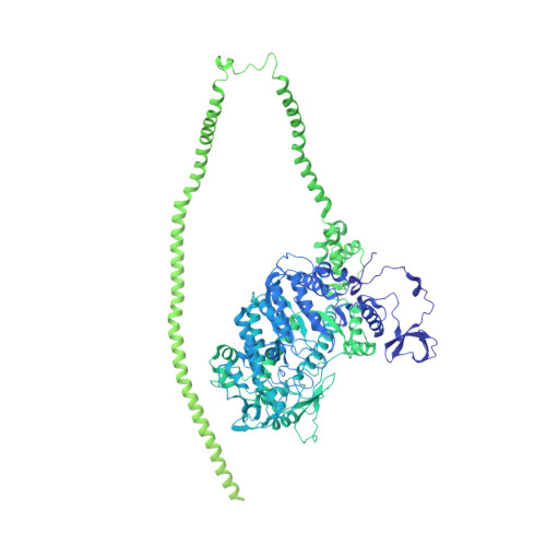
Myosin-7
A, B
1,935Homo sapiensMutation(s): 0 
UniProt & NIH Common Fund Data Resources
Find proteins for P12883 (Homo sapiens)
Explore P12883 
Go to UniProtKB:  P12883
PHAROS:  P12883
GTEx:  ENSG00000092054 
Entity Groups  
Sequence Clusters30% Identity50% Identity70% Identity90% Identity95% Identity100% Identity
UniProt GroupP12883
Sequence Annotations
Expand
  • Reference Sequence
Find similar proteins by:  (by identity cutoff)  |  3D Structure
Entity ID: 2
MoleculeChains Sequence LengthOrganismDetailsImage
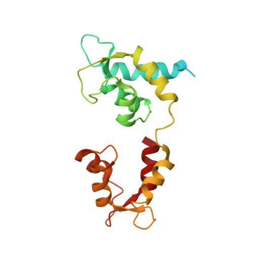
Myosin light chain 3
C, D
195Homo sapiensMutation(s): 0 
UniProt & NIH Common Fund Data Resources
Find proteins for P08590 (Homo sapiens)
Explore P08590 
Go to UniProtKB:  P08590
PHAROS:  P08590
GTEx:  ENSG00000160808 
Entity Groups  
Sequence Clusters30% Identity50% Identity70% Identity90% Identity95% Identity100% Identity
UniProt GroupP08590
Sequence Annotations
Expand
  • Reference Sequence
Find similar proteins by:  (by identity cutoff)  |  3D Structure
Entity ID: 3
MoleculeChains Sequence LengthOrganismDetailsImage
Myosin regulatory light chain 2, ventricular/cardiac muscle isoform
E, F
166Homo sapiensMutation(s): 0 
UniProt & NIH Common Fund Data Resources
Find proteins for P10916 (Homo sapiens)
Explore P10916 
Go to UniProtKB:  P10916
PHAROS:  P10916
GTEx:  ENSG00000111245 
Entity Groups  
Sequence Clusters30% Identity50% Identity70% Identity90% Identity95% Identity100% Identity
UniProt GroupP10916
Sequence Annotations
Expand
  • Reference Sequence
Experimental Data & Validation

Experimental Data

  • Method: ELECTRON MICROSCOPY
  • Resolution: 20.0 Å
  • Aggregation State: FILAMENT 
  • Reconstruction Method: SINGLE PARTICLE 
EM Software:
TaskSoftware PackageVersion
RECONSTRUCTIONITERATIVE HELICAL REAL SPACE RECONSTRUCTION (EGELMAN, 2000)SPIDER 14

Structure Validation

View Full Validation Report


Entry History & Funding Information

Deposition Data


Funding OrganizationLocationGrant Number
Howard Hughes Medical Institute (HHMI)United States--
Leducq FoundationFrance--
Wellcome TrustUnited States--
National Institutes of Health/National Heart, Lung, and Blood Institute (NIH/NHLBI)United StatesNHLBI-HL084553
National Science Foundation (NSF, United States)United StatesDMR-1332208
National Institutes of Health/National Institute of General Medical Sciences (NIH/NIGMS)United StatesGM-103485
Medical Research Council (MRC, United Kingdom)United Kingdom--

Revision History  (Full details and data files)

  • Version 1.0: 2017-06-07
    Type: Initial release
  • Version 1.1: 2017-06-28
    Changes: Database references
  • Version 1.2: 2017-08-09
    Changes: Database references
  • Version 1.3: 2017-09-20
    Changes: Author supporting evidence
  • Version 1.4: 2017-11-08
    Changes: Derived calculations
  • Version 1.5: 2018-07-18
    Changes: Data collection
  • Version 1.6: 2018-10-03
    Changes: Data collection, Refinement description
  • Version 1.7: 2019-11-20
    Changes: Author supporting evidence
  • Version 1.8: 2020-01-08
    Changes: Author supporting evidence
  • Version 1.9: 2024-10-23
    Changes: Data collection, Database references, Derived calculations, Refinement description, Structure summary