5KP7 | pdb_00005kp7

Crystal Structure of the Curacin Biosynthetic Pathway HMG Synthase in Complex with Holo Donor-ACP


Experimental Data Snapshot

  • Method: X-RAY DIFFRACTION
  • Resolution: 1.60 Å
  • R-Value Free: 
    0.167 (Depositor), 0.170 (DCC) 
  • R-Value Work: 
    0.151 (Depositor), 0.160 (DCC) 
  • R-Value Observed: 
    0.152 (Depositor) 

Starting Models: experimental
View more details

wwPDB Validation   3D Report Full Report


Ligand Structure Quality Assessment 

Created with Raphaël 2.3.0Worse 01 BetterLigand structure goodness of fit to experimental dataBest fitted PNSClick on this verticalbar to view details

This is version 1.5 of the entry. See complete history


Literature

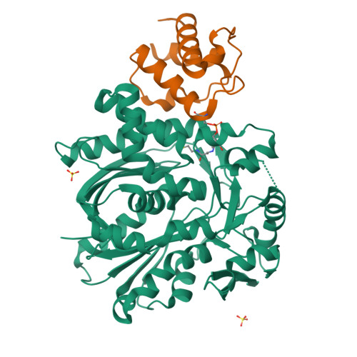
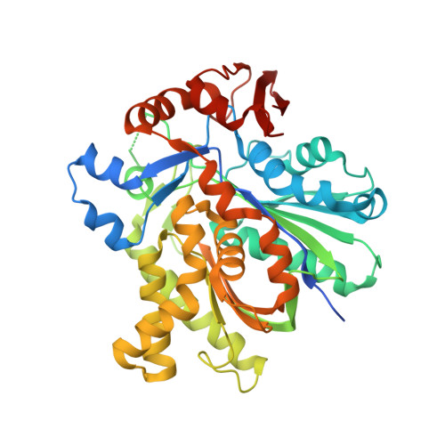
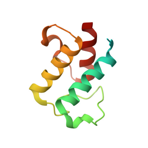
Anatomy of the beta-branching enzyme of polyketide biosynthesis and its interaction with an acyl-ACP substrate.

Maloney, F.P.Gerwick, L.Gerwick, W.H.Sherman, D.H.Smith, J.L.

(2016) Proc Natl Acad Sci U S A 113: 10316-10321

  • DOI: https://doi.org/10.1073/pnas.1607210113
  • Primary Citation of Related Structures:  
    5KP5, 5KP6, 5KP7, 5KP8

  • PubMed Abstract: 

    Alkyl branching at the β position of a polyketide intermediate is an important variation on canonical polyketide natural product biosynthesis. The branching enzyme, 3-hydroxy-3-methylglutaryl synthase (HMGS), catalyzes the aldol addition of an acyl donor to a β-keto-polyketide intermediate acceptor. HMGS is highly selective for two specialized acyl carrier proteins (ACPs) that deliver the donor and acceptor substrates. The HMGS from the curacin A biosynthetic pathway (CurD) was examined to establish the basis for ACP selectivity. The donor ACP (CurB) had high affinity for the enzyme (Kd = 0.5 μM) and could not be substituted by the acceptor ACP. High-resolution crystal structures of HMGS alone and in complex with its donor ACP reveal a tight interaction that depends on exquisite surface shape and charge complementarity between the proteins. Selectivity is explained by HMGS binding to an unusual surface cleft on the donor ACP, in a manner that would exclude the acceptor ACP. Within the active site, HMGS discriminates between pre- and postreaction states of the donor ACP. The free phosphopantetheine (Ppant) cofactor of ACP occupies a conserved pocket that excludes the acetyl-Ppant substrate. In comparison with HMG-CoA (CoA) synthase, the homologous enzyme from primary metabolism, HMGS has several differences at the active site entrance, including a flexible-loop insertion, which may account for the specificity of one enzyme for substrates delivered by ACP and the other by CoA.


  • Organizational Affiliation

    Life Sciences Institute, University of Michigan, Ann Arbor, MI 48109; Chemical Biology Doctoral Program, University of Michigan, Ann Arbor, MI 48109;


Macromolecules
Find similar proteins by:  (by identity cutoff)  |  3D Structure
Entity ID: 1
MoleculeChains Sequence LengthOrganismDetailsImage
CurD443Moorena producens 3LMutation(s): 3 
Gene Names: LYNGBM3L_74540
EC: 2.3.3.10
UniProt
Find proteins for F4Y432 (Moorena producens 3L)
Explore F4Y432 
Go to UniProtKB:  F4Y432
Entity Groups  
Sequence Clusters30% Identity50% Identity70% Identity90% Identity95% Identity100% Identity
UniProt GroupF4Y432
Sequence Annotations
Expand
  • Reference Sequence
Find similar proteins by:  (by identity cutoff)  |  3D Structure
Entity ID: 2
MoleculeChains Sequence LengthOrganismDetailsImage
CurB103Lyngbya majusculaMutation(s): 0 
Gene Names: curB
UniProt
Find proteins for Q6DNF1 (Lyngbya majuscula)
Explore Q6DNF1 
Go to UniProtKB:  Q6DNF1
Entity Groups  
Sequence Clusters30% Identity50% Identity70% Identity90% Identity95% Identity100% Identity
UniProt GroupQ6DNF1
Sequence Annotations
Expand
  • Reference Sequence
Experimental Data & Validation

Experimental Data

  • Method: X-RAY DIFFRACTION
  • Resolution: 1.60 Å
  • R-Value Free:  0.167 (Depositor), 0.170 (DCC) 
  • R-Value Work:  0.151 (Depositor), 0.160 (DCC) 
  • R-Value Observed: 0.152 (Depositor) 
Space Group: P 31 2 1
Unit Cell:
Length ( Å )Angle ( ˚ )
a = 101.117α = 90
b = 101.117β = 90
c = 104.595γ = 120
Software Package:
Software NamePurpose
REFMACrefinement
PDB_EXTRACTdata extraction
XDSdata reduction
XDSdata scaling
REFMACphasing

Structure Validation

View Full Validation Report


Ligand Structure Quality Assessment 

Created with Raphaël 2.3.0Worse 01 BetterLigand structure goodness of fit to experimental dataBest fitted PNSClick on this verticalbar to view details

Entry History & Funding Information

Deposition Data


Funding OrganizationLocationGrant Number
National Institutes of Health/National Institute of Diabetes and Digestive and Kidney Disease (NIH/NIDDK)United StatesR01-DK042303
National Institutes of Health/National Cancer Institute (NIH/NCI)United StatesR01-CA108874

Revision History  (Full details and data files)

  • Version 1.0: 2016-08-31
    Type: Initial release
  • Version 1.1: 2016-09-14
    Changes: Database references
  • Version 1.2: 2016-09-21
    Changes: Database references
  • Version 1.3: 2017-09-20
    Changes: Author supporting evidence, Database references, Derived calculations
  • Version 1.4: 2019-12-04
    Changes: Author supporting evidence
  • Version 1.5: 2023-10-04
    Changes: Data collection, Database references, Derived calculations, Refinement description