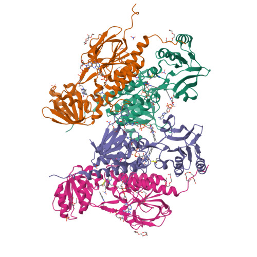
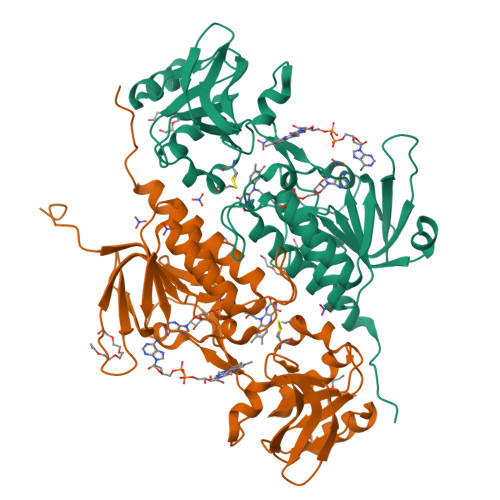
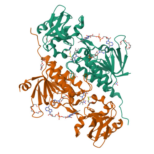
5K0A

Structure of an oxidoreductase from Synechocystis sp. PCC6803


Experimental Data Snapshot

  • Method: X-RAY DIFFRACTION
  • Resolution: 1.71 Å
  • R-Value Free: 0.193 
  • R-Value Work: 0.165 
  • R-Value Observed: 0.166 

Starting Model: experimental
View more details

wwPDB Validation   3D Report Full Report


Ligand Structure Quality Assessment 


This is version 1.5 of the entry. See complete history


Literature

Unprecedented pathway of reducing equivalents in a diflavin-linked disulfide oxidoreductase.

Buey, R.M.Arellano, J.B.Lopez-Maury, L.Galindo-Trigo, S.Velazquez-Campoy, A.Revuelta, J.L.de Pereda, J.M.Florencio, F.J.Schurmann, P.Buchanan, B.B.Balsera, M.

(2017) Proc Natl Acad Sci U S A 114: 12725-12730

  • DOI: https://doi.org/10.1073/pnas.1713698114
  • Primary Citation of Related Structures:  
    5JRI, 5K0A, 5N0J, 5ODE

  • PubMed Abstract: 

    Flavoproteins participate in a wide variety of physiologically relevant processes that typically involve redox reactions. Within this protein superfamily, there exists a group that is able to transfer reducing equivalents from FAD to a redox-active disulfide bridge, which further reduces disulfide bridges in target proteins to regulate their structure and function. We have identified a previously undescribed type of flavin enzyme that is exclusive to oxygenic photosynthetic prokaryotes and that is based on the primary sequence that had been assigned as an NADPH-dependent thioredoxin reductase (NTR). However, our experimental data show that the protein does not transfer reducing equivalents from flavins to disulfides as in NTRs but functions in the opposite direction. High-resolution structures of the protein from Gloeobacter violaceus and Synechocystis sp. PCC6803 obtained by X-ray crystallography showed two juxtaposed FAD molecules per monomer in redox communication with an active disulfide bridge in a variant of the fold adopted by NTRs. We have tentatively named the flavoprotein "DDOR" (diflavin-linked disulfide oxidoreductase) and propose that its activity is linked to a thiol-based transfer of reducing equivalents in bacterial membranes. These findings expand the structural and mechanistic repertoire of flavoenzymes with oxidoreductase activity and pave the way to explore new protein engineering approaches aimed at designing redox-active proteins for diverse biotechnological applications.


  • Organizational Affiliation

    Metabolic Engineering Group, Departamento de Microbiología y Genética, Universidad de Salamanca, 37007 Salamanca, Spain.


Macromolecules
Find similar proteins by:  (by identity cutoff)  |  3D Structure
Entity ID: 1
MoleculeChains Sequence LengthOrganismDetailsImage
Slr0600 protein
A, B, C, D
330Synechocystis sp. PCC 6803 substr. KazusaMutation(s): 0 
Gene Names: slr0600
UniProt
Find proteins for P74746 (Synechocystis sp. (strain ATCC 27184 / PCC 6803 / Kazusa))
Explore P74746 
Go to UniProtKB:  P74746
Entity Groups  
Sequence Clusters30% Identity50% Identity70% Identity90% Identity95% Identity100% Identity
UniProt GroupP74746
Sequence Annotations
Expand
  • Reference Sequence
Small Molecules
Ligands 6 Unique
IDChains Name / Formula / InChI Key2D Diagram3D Interactions
FAD
Query on FAD

Download Ideal Coordinates CCD File 
BA [auth D]
CA [auth D]
E [auth A]
F [auth A]
M [auth B]
BA [auth D],
CA [auth D],
E [auth A],
F [auth A],
M [auth B],
N [auth B],
U [auth C],
V [auth C]
FLAVIN-ADENINE DINUCLEOTIDE
C27 H33 N9 O15 P2
VWWQXMAJTJZDQX-UYBVJOGSSA-N
1PE
Query on 1PE

Download Ideal Coordinates CCD File 
AA [auth D],
GA [auth D],
P [auth B],
Y [auth C],
Z [auth C]
PENTAETHYLENE GLYCOL
C10 H22 O6
JLFNLZLINWHATN-UHFFFAOYSA-N
PG4
Query on PG4

Download Ideal Coordinates CCD File 
FA [auth D]
I [auth A]
J [auth A]
R [auth B]
S [auth B]
FA [auth D],
I [auth A],
J [auth A],
R [auth B],
S [auth B],
W [auth C]
TETRAETHYLENE GLYCOL
C8 H18 O5
UWHCKJMYHZGTIT-UHFFFAOYSA-N
PGE
Query on PGE

Download Ideal Coordinates CCD File 
HA [auth D]TRIETHYLENE GLYCOL
C6 H14 O4
ZIBGPFATKBEMQZ-UHFFFAOYSA-N
PO4
Query on PO4

Download Ideal Coordinates CCD File 
DA [auth D],
T [auth B]
PHOSPHATE ION
O4 P
NBIIXXVUZAFLBC-UHFFFAOYSA-K
NO3
Query on NO3

Download Ideal Coordinates CCD File 
EA [auth D]
G [auth A]
H [auth A]
K [auth A]
L [auth A]
EA [auth D],
G [auth A],
H [auth A],
K [auth A],
L [auth A],
O [auth B],
Q [auth B],
X [auth C]
NITRATE ION
N O3
NHNBFGGVMKEFGY-UHFFFAOYSA-N
Modified Residues  1 Unique
IDChains TypeFormula2D DiagramParent
CSO
Query on CSO
A, B, C, D
L-PEPTIDE LINKINGC3 H7 N O3 SCYS
Experimental Data & Validation

Experimental Data

  • Method: X-RAY DIFFRACTION
  • Resolution: 1.71 Å
  • R-Value Free: 0.193 
  • R-Value Work: 0.165 
  • R-Value Observed: 0.166 
  • Space Group: P 1 21 1
Unit Cell:
Length ( Å )Angle ( ˚ )
a = 66.029α = 90
b = 128.43β = 91.61
c = 92.728γ = 90
Software Package:
Software NamePurpose
PHENIXrefinement
XDSdata reduction
XSCALEdata scaling
PHASERphasing

Structure Validation

View Full Validation Report


Ligand Structure Quality Assessment 


Entry History 

Deposition Data

Revision History  (Full details and data files)

  • Version 1.0: 2017-11-15
    Type: Initial release
  • Version 1.1: 2017-11-22
    Changes: Database references
  • Version 1.2: 2017-12-13
    Changes: Database references
  • Version 1.3: 2021-09-08
    Changes: Database references
  • Version 1.4: 2024-01-10
    Changes: Data collection, Refinement description
  • Version 1.5: 2024-10-23
    Changes: Structure summary